Image processing method, device, electronic device and storage medium
An image processing and image technology, applied in the field of medical image processing, can solve the problems of increasing the scanning time, excessive radiation dose to the patient, etc., to reduce the radiation dose, improve the probability of information retention, and enhance the effect of detail information.
- Summary
- Abstract
- Description
- Claims
- Application Information
AI Technical Summary
Problems solved by technology
Method used
Image

Examples
Embodiment 1
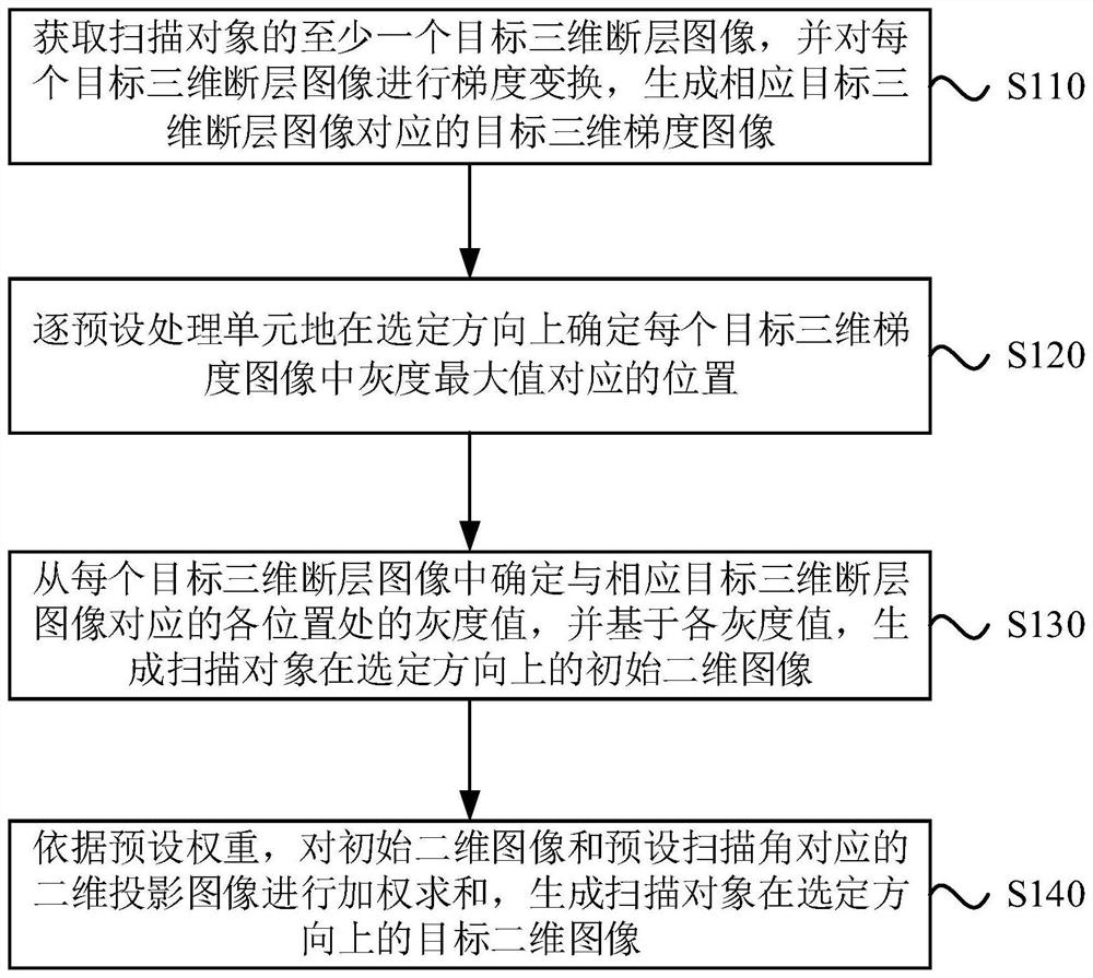
[0031] The image processing method provided in this embodiment is applicable to acquiring a two-dimensional image in a selected direction from a three-dimensional image. The method can be executed by an image processing device, which can be implemented by software and / or hardware, and which can be integrated in electronic equipment with image processing functions, such as personal computers, servers or network equipment. see figure 1 , the method of this embodiment specifically includes the following steps:
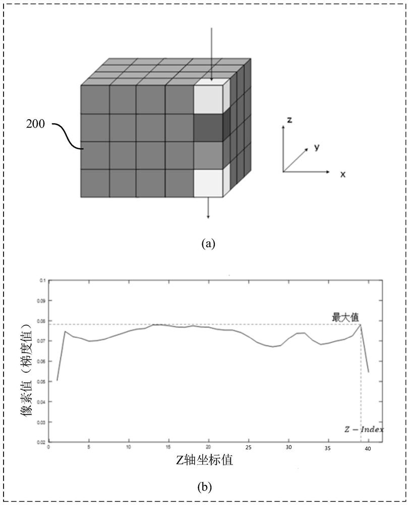
[0032] S110. Acquire at least one target three-dimensional tomographic image of the scanned object, and perform gradient transformation on each target three-dimensional tomographic image to generate a target three-dimensional gradient image corresponding to the corresponding target three-dimensional tomographic image.
[0033] Wherein, the target 3D tomographic image refers to a 3D tomographic image that can be directly used to extract a 2D image. The three-dimensional ...
Embodiment 2
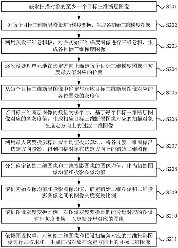
[0058] In this embodiment, on the basis of the above-mentioned first embodiment, “gradient transformation is performed on each target 3D tomographic image to generate a target gradient image corresponding to the corresponding target 3D tomographic image” is further optimized. On this basis, it is also possible to further optimize "project each transitional two-dimensional image along the selected direction to generate the initial two-dimensional image of the scanned object in the selected direction". On the basis of the above, a step of gray scale normalization between the "initial two-dimensional image and the two-dimensional projected image" can be further added. The number of target three-dimensional tomographic images in this embodiment is multiple. The explanations of terms that are the same as or corresponding to the above-mentioned embodiments will not be repeated here. see image 3 , the image processing method provided in this embodiment includes:
[0059] S201. Ac...
Embodiment 3
[0082] On the basis of the above-mentioned embodiments, this embodiment further optimizes "acquiring at least one target three-dimensional tomographic image of the scanning object". On this basis, the step of enhancing and correcting the target two-dimensional image may be further added. The explanations of terms that are the same as or corresponding to the above-mentioned embodiments will not be repeated here. see Figure 5 , the image processing method provided in this embodiment includes:
[0083] S301. Acquire at least one initial three-dimensional tomographic image of a scanned object.
[0084] The initial three-dimensional tomographic image is an image obtained by three-dimensional reconstruction after scanning the scanned object, which does not undergo further image post-processing operations. The initial three-dimensional tomographic image can be obtained by scanning the scanning part of the scanning object in real time, or can be obtained by reading from an externa...
PUM
 Abstract
Description
Claims
Application Information
 - R&D
- Intellectual Property
- Life Sciences
- Materials
- Tech Scout
- Unparalleled Data Quality
- Higher Quality Content
- 60% Fewer Hallucinations
Browse by: Latest US Patents, China's latest patents, Technical Efficacy Thesaurus, Application Domain, Technology Topic, Popular Technical Reports.
© 2025 PatSnap. All rights reserved.Legal|Privacy policy|Modern Slavery Act Transparency Statement|Sitemap|About US| Contact US: help@patsnap.com