CT image liver tumor segmentation method based on deep learning

A CT image, liver tumor technology, applied in the field of medical image processing, can solve the problem of no fully connected layer, and achieve the effect of improving efficiency and accuracy

Pending Publication Date: 2019-12-13
BEIJING UNIV OF TECH
View PDF1 Cites 7 Cited by
  • Summary
  • Abstract
  • Description
  • Claims
  • Application Information

AI Technical Summary

Problems solved by technology

"Fully Convolutional Networks for Semantic Segmentation.", Long, Jonathan, Shelhamer, Evan and Darrell, Trevor, The IEEE Conference on ComputerVision and Pattern Recognition (CVPR

Method used


Image

  • CT image liver tumor segmentation method based on deep learning
  • CT image liver tumor segmentation method based on deep learning
  • CT image liver tumor segmentation method based on deep learning

Examples


Example Embodiment

[0016] Specific implementation method

[0017] The present invention will be further explained below in conjunction with examples

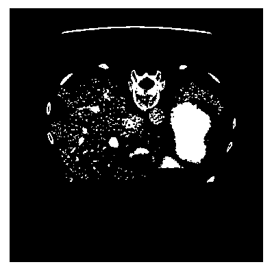
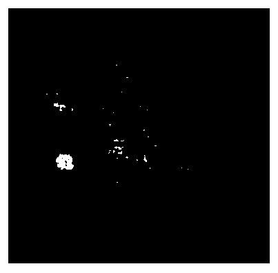
[0018] The present invention provides a CT image liver tumor segmentation method based on deep learning, using a data set from the LiTS (Liver Tumor Segmentation Challenge, CT image segmentation challenge of liver tumor lesions) data set, and LiTS is a data set for liver tumor segmentation. It contains 131 sets of training data and 70 sets of test data. The training data contains 131 sets of 3D CT images and corresponding 131 sets of real segmentation masks.

[0019] Before using the data, it is necessary to preprocess the CT image data, first convert the CT value of the CT image to the HU value. The scope of the data is limited. In this experiment, the HU value of the atlas is set to include but not limited to [-200, 250], and some irrelevant information and noise are removed. Divide the ROI for cropping, divide the area of ​​the liver, and perform c...


PUM


Abstract

The invention relates to a CT image liver tumor segmentation method based on deep learning, and specifically solves the problems that the original U-Net depth is small, the structure is simple, a model with a good training effect is difficult to train, and the accuracy of CT image liver tumor segmentation is insufficient. Specifically, the network structure of the U-Net is optimized and improved;a Dropout layer and a Batch Normalization layer are added to modify a network structure, a VGG-16 network module is used to replace an encoder part of U-Net, and an optimized model is established to perform accurate segmentation on a liver tumor CT image.

Description

technical field [0001] The invention belongs to the field of medical image processing, and relates to a method for segmenting liver tumors in CT images based on deep learning. Background technique [0002] The liver is the largest solid organ in the abdominal cavity of the human body, and it has a very complex structure and abundant blood vessels. And the liver has many types of lesions and a high incidence rate. The precise segmentation of liver tumor images plays a vital role in the treatment of the liver, but due to the complexity of the size, shape, and location of liver tumors, its segmentation is difficult and computationally intensive for traditional machine learning methods . With the rapid development of deep learning in the field of machine vision and the rapid growth of the number of medical images, medical image analysis based on deep learning has gradually become an important auxiliary tool for treatment. [0003] Many researchers have done a lot of research ...

Claims


Application Information

Patent Timeline
IPC IPC(8): G06T7/11G06N3/04G06N3/08
CPCG06T7/11G06N3/08G06T2207/10081G06T2207/30056G06N3/045
Inventor 王瑾熊志琪朱青
Owner BEIJING UNIV OF TECH
Why Eureka
Try Eureka