Method and presentation device for post processing and displaying a three-dimensional angiography image data set

An image data set, a technology for displaying images, applied in image data processing, 3D image processing, medical images, etc., to improve diagnosis, improve three-dimensional perception, and speed up workflow

Pending Publication Date: 2020-01-07
SIEMENS HEALTHCARE GMBH
View PDF6 Cites 0 Cited by
  • Summary
  • Abstract
  • Description
  • Claims
  • Application Information

AI Technical Summary

Problems solved by technology

However, this visualization requires a high degree of spatial awareness on the user's side

Method used


Image

  • Method and presentation device for post processing and displaying a three-dimensional angiography image data set
  • Method and presentation device for post processing and displaying a three-dimensional angiography image data set
  • Method and presentation device for post processing and displaying a three-dimensional angiography image data set

Examples


Embodiment Construction

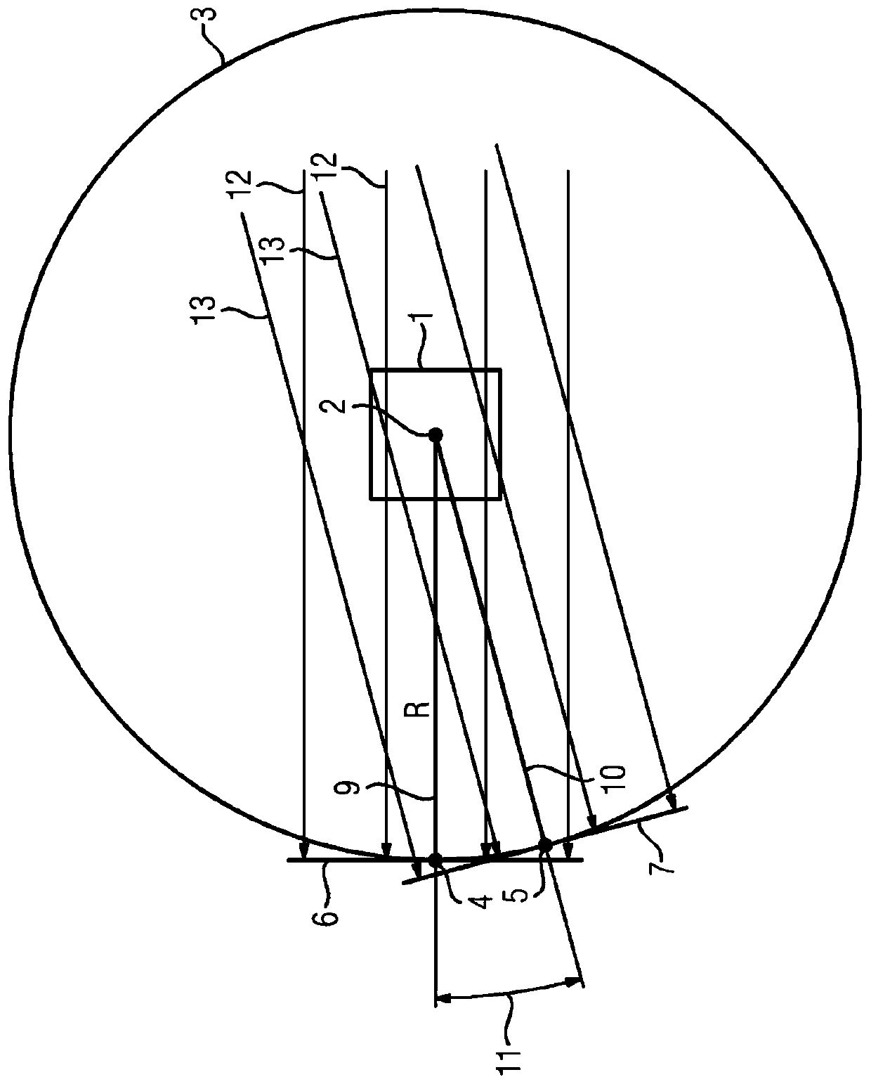
[0032] The following embodiments exemplarily relate to a three-dimensional magnetic resonance angiography image data set acquired with a magnetic resonance apparatus, however, the embodiments can also be applied to other imaging techniques as three-dimensional data sources. Embodiments described herein describe the visualization (i.e., rendering) of a vascular tree described by an angiographic image dataset from at least one mean viewing direction, where, in general, as a rendering technique, parallel maximum intensity projection ( MIP) to calculate the two display images for simultaneous display. The viewing directions of the pairs of display images to be displayed simultaneously (each direction to a different eye of the user) draw an angle less than 10° (eg, 5°) suitable for stereoscopic viewing.

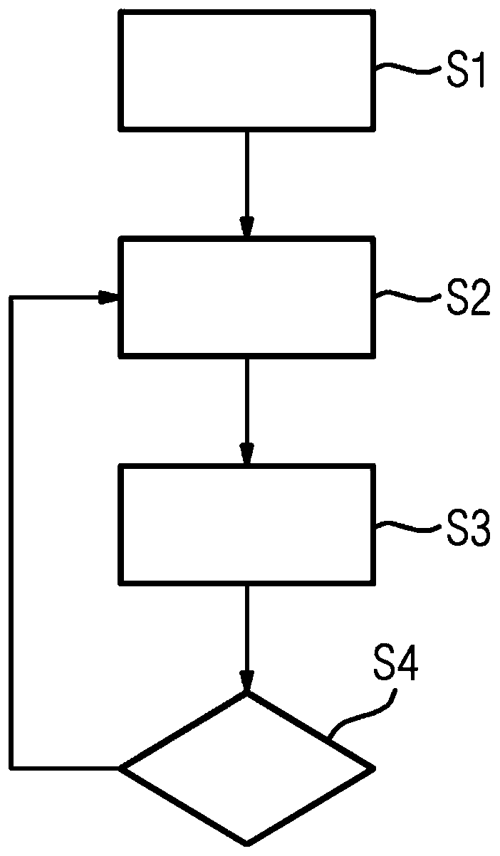
[0033] figure 1 A flow diagram of an embodiment of the method according to the invention is shown. In step S1, a rendering geometry for rendering a display image is defined. Fo...


PUM


Abstract

The invention provides a method and a presentation device for post processing and displaying a three-dimensional angiography image data set. The invention provides a method for post processing and displaying a three-dimensional angiography image data set of a blood vessel tree of a patient, wherein two-dimensional display images are rendered from the angiography image data set and displayed, wherein two display images are rendered from the angiography image data set using viewing directions (9, 10) forming an angle (11) suited for stereoscopic perception of the display images and both displayimages are simultaneously displayed on a display device (15) such that each display image is displayed to one eye of a user.

Description

technical field [0001] The invention relates to a method for postprocessing and displaying a three-dimensional angiographic image data set of a vascular tree of a patient, wherein a two-dimensional display image is rendered from the angiographic image data set and displayed. The invention also relates to a presentation device, a computer program and an electronically readable storage medium. Background technique [0002] Angiography is a well known medical imaging technique used to visualize blood vessels in a patient. A three-dimensional angiographic image dataset may be acquired using an x-ray imaging device, in which a contrast agent is injected into the patient to allow blood vessels to be contrasted with other anatomical structures. However, it has also been proposed to use Magnetic Resonance Imaging (MRI) to image blood vessels of a patient, eg to diagnose stenosis, occlusion or aneurysm. In any case, the result of the acquisition is a three-dimensional angiographic ...

Claims


Application Information

Patent Timeline
Patent Type & Authority Applications(China)
IPC IPC(8): G16H30/40G06T15/20G06T15/50
CPCG16H30/40G06T15/205G06T15/50H04N13/275G06T15/08A61B6/466A61B6/504A61B8/466H04N13/344H04N13/351G06T11/008G06T2210/41G06T2211/404
Inventor 朱利安·霍斯巴赫雷纳·施奈德彼得·施派尔
Owner SIEMENS HEALTHCARE GMBH
Why Eureka
Try Eureka