Immunohistochemical membrane staining section diagnosis method and device

A technology of immunohistochemistry and diagnostic methods, which is applied in the field of immunohistochemical membrane staining section diagnostic methods and devices, which can solve the problems of cell number statistics, cell type discrimination, and different staining effects, and achieve robustness. Good performance, guaranteed accuracy, and convenient process operation

Pending Publication Date: 2020-02-28
杭州迪英加科技有限公司
View PDF5 Cites 16 Cited by
  • Summary
  • Abstract
  • Description
  • Claims
  • Application Information

AI Technical Summary

Problems solved by technology

[0003] The purpose of the present invention is to solve the technical problem that the immunohistochemical membrane staining sections in the prior art are easy to affect the statistics of cell numbers and the discrimination of cell types due to the different staining effects. The present invention provides an immunohistochemical membrane Diagnosis method and device for membrane stained section

Method used


Image

  • Immunohistochemical membrane staining section diagnosis method and device
  • Immunohistochemical membrane staining section diagnosis method and device
  • Immunohistochemical membrane staining section diagnosis method and device

Examples


Embodiment Construction

[0021] The present invention will be further described below in conjunction with accompanying drawing:

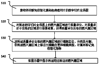
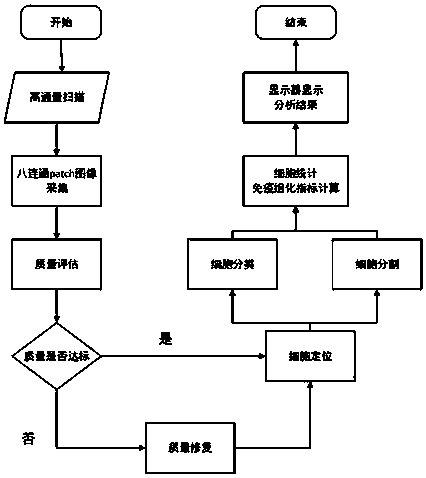
[0022] A diagnostic device for immunohistochemical membrane staining slices, including a high-throughput scanner, a processor and a display. The processor is communicated with the high-throughput scanner, and the processor is connected with the display. Immunohistochemical membrane stained pathological sections are placed under a high-throughput scanner, and the high-throughput scanner scans immunohistochemical membrane-stained pathological sections into digital slice full-field images, which are sent to the processor for processing, and the digital images processed by the processor The sliced ​​full-field image output is displayed on the monitor. The high-throughput scanner used in this example is used to scan immunohistochemically stained sections and perform digital visualization of the sections, including magnifications of 4×, 10× and 40×.

[0023] Such as figure 1 ,...


PUM


Abstract

The invention relates to an immunohistochemical membrane staining section diagnosis method. The method comprises the following steps: S10, receiving a digital section full-field graph of a to-be-diagnosed immunohistochemical membrane staining pathological section; S20, performing quality evaluation on the region of interest on the digital slice full-field image, and performing image restoration onthe region with unqualified quality evaluation to obtain a region of interest with qualified quality evaluation; S30, carrying out cell positioning, classification and segmentation on the regions ofinterest with qualified quality evaluation, marking cell positions and cell types on the regions of interest, and calculating and marking immunohistochemical indexes; and S40, displaying the marked region of interest. The invention further relates to an immunohistochemical membrane staining section diagnosis device and a computer readable medium. Since the context information of the lesion area isof great significance to interpretation of the immunohistochemical membrane staining section, the accuracy of interpretation is ensured by collecting the eight-connected patch image of the immunohistochemical membrane staining pathological section with time sequence for training and testing.

Description

technical field [0001] The invention relates to the field of section diagnosis, in particular to a method and device for immunohistochemical membrane staining section diagnosis. Background technique [0002] As a key technology of histopathology or cytopathology, immunohistochemistry technology is one of the key technologies in the current process of tumor interpretation, diagnosis and treatment, and has great influence. The so-called immunohistochemical technique or immunocytochemical technique refers to the qualitative, localization or quantitative study of certain chemical components in tissue sections or cell specimens by applying the principles of immunology and histochemistry. Immunohistochemical histopathology techniques are divided into immunohistochemical membrane staining and plasma cell staining. For immunohistochemical membrane staining sections, the staining process is done manually by staining technicians. Because staining technicians have different control ove...

Claims


Application Information

Patent Timeline
Patent Type & Authority Applications(China)
IPC IPC(8): G06T7/00G06T7/11G06T7/70G06K9/32
CPCG06T7/0012G06T7/70G06T7/11G06T2207/30024G06T2207/30096G06T2207/30204G06T2207/20104G06V10/25
Inventor 亢宇鑫林明珍崔灿崔磊杨林彭之磊
Owner 杭州迪英加科技有限公司
Why Eureka
Try Eureka