
Position classification method, device and equipment for pulmonary nodules and storage medium

A technology for pulmonary nodules and nodules, which is applied in the classification method of pulmonary nodules, equipment and storage media, and devices, can solve the problems of not being intuitive and clear enough, reducing workload and screening, and displaying complex results, so as to achieve an improvement Diagnosis efficiency, position results are intuitive and clear, and the effect of reducing workload

Active Publication Date: 2020-03-24
SHANGHAI UNITED IMAGING INTELLIGENT MEDICAL TECH CO LTD
View PDF4 Cites 4 Cited by
  • Summary
  • Abstract
  • Description
  • Claims
  • Application Information

AI Technical Summary

Problems solved by technology

[0004] In the research of the prior art, the classification of pulmonary nodules is mainly aimed at the classification of the lesion types of pulmonary nodules, and at the same time, the positioning function of pulmonary nodules can be realized, and finally The specific location of each pulmonary nodule can be displayed on the report, but this method is not intuitive and clear when there are many pulmonary nodules displayed, and the display results are more complicated, which cannot better help doctors reduce workload and screening. Therefore, a more efficient solution is required

Method used


Image

  • Position classification method, device and equipment for pulmonary nodules and storage medium
  • Position classification method, device and equipment for pulmonary nodules and storage medium
  • Position classification method, device and equipment for pulmonary nodules and storage medium

Examples


Embodiment Construction

[0037] The following will clearly and completely describe the technical solutions in the embodiments of the application with reference to the drawings in the embodiments of the application. Apparently, the described embodiments are only some of the embodiments of the application, not all of them. All other embodiments obtained by persons of ordinary skill in the art based on the embodiments in the present application without creative efforts shall fall within the protection scope of the present application. Examples of the described embodiments are shown in the drawings, wherein like or similar reference numerals designate like or similar elements or elements having the same or similar functions throughout.

[0038] It should be noted that the terms "first" and "second" in the description and claims of the present application and the above drawings are used to distinguish similar objects, but not necessarily used to describe a specific sequence or sequence. It is to be underst...


PUM


Abstract

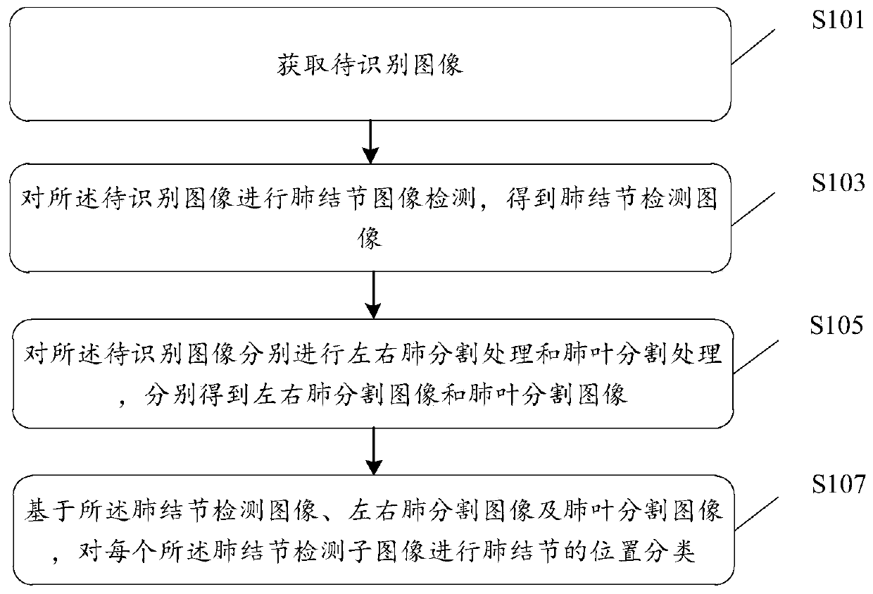
The invention discloses a position classification method, device and equipment for pulmonary nodules, and a storage medium. The method comprises the steps of obtaining a to-be-identified image; carrying out pulmonary nodule image detection on the to-be-identified image to obtain a pulmonary nodule detection image, wherein the pulmonary nodule detection image comprises one or more pulmonary noduledetection sub-images; performing left and right lung segmentation processing and lung lobe segmentation processing on the to-be-identified image to obtain left and right lung segmentation images and alung lobe segmentation image; and based on the pulmonary nodule detection image, the left and right lung segmentation images and the lung lobe segmentation image, performing pulmonary nodule positionclassification on each pulmonary nodule detection sub-image. By means of the technical scheme, lung morphological characteristics can be combined, pulmonary nodule position classification is assistedthrough left and right lung segmentation and lung lobe segmentation, the classification speed can be increased, the obtained pulmonary nodule position result is more intuitive and clearer, and follow-up screening for pulmonary nodules of different position types is facilitated.

Description

technical field [0001] The present invention relates to the technical field of image processing, in particular to a location classification method, device, equipment and storage medium for pulmonary nodules. Background technique [0002] With the continuous improvement of the medical level, people will use various medical images to assist the diagnosis in the diagnosis process. Computed tomography (CT) is the most commonly used technology. Doctors usually make a diagnosis by looking at the CT image data of the lungs, but The distribution of pulmonary nodules is indeterminate, and it is very easy to connect with other tissues. Therefore, doctors have a large workload in diagnosis, and misdiagnosis and missed diagnosis are prone to occur. Even for experienced doctors, under the burden of a large amount of image data, it is difficult to make an objective and accurate judgment all the time, which eventually leads to the delay of the best treatment time for patients. Therefore, i...

Claims


Application Information

Patent Timeline
IPC IPC(8): G06T7/00G06T7/11G06K9/62
CPCG06T7/0012G06T7/11G06T2207/10081G06T2207/20081G06T2207/20084G06T2207/30064G06F18/241
Inventor 张文海高耀宗
Owner SHANGHAI UNITED IMAGING INTELLIGENT MEDICAL TECH CO LTD
Learn More