A surface-enhanced Raman scattering substrate material for gas detection and its preparation method

A surface-enhanced Raman and gas detection technology, used in Raman scattering, material analysis, material excitation analysis, etc. Effect

Active Publication Date: 2022-03-25
WENZHOU INST UNIV OF CHINESE ACAD OF SCI
View PDF6 Cites 0 Cited by
  • Summary
  • Abstract
  • Description
  • Claims
  • Application Information

AI Technical Summary

Problems solved by technology

However, this method has high specificity, and can only specifically detect a certain kind or a certain type of gas molecules, such as the preparation method of the surface-enhanced Raman scattering substrate material and the gas detection method of the patent No. CN108072641A, through Functional modification of one or more of 4-aminothiophenol, 4-hydroxythiophenol or 2-carboxythiophenol, preferably 4-aminothiophenol, the SERS substrate material prepared by the above method can only target Single type of gas detection, no response to other gases, so the scope of application is limited

Method used


Image

  • A surface-enhanced Raman scattering substrate material for gas detection and its preparation method
  • A surface-enhanced Raman scattering substrate material for gas detection and its preparation method
  • A surface-enhanced Raman scattering substrate material for gas detection and its preparation method

Examples


Embodiment 1

[0033] A method for preparing a surface-enhanced Raman scattering base material for gas detection, specifically comprising the following steps:

[0034] Step 1. Cut the glass into small squares of 0.6×0.6cm, grind the base with sandpaper to form a rough working surface, and then use acetone, isopropanol, and ethanol for ultrasonic cleaning in sequence, each ultrasonic cleaning time is 20-30min, and finally dry the glass with nitrogen piece;

[0035] Step 2. Soak the substrate prepared in step 1 in 10mL of 10mM tetrachloroauric acid trihydrate aqueous solution, add ammonia water with a concentration of 0.6%, control pH 8-10, and oscillate at an oscillation frequency of 500rpm for 5min to make chloroauric acid Ions are adsorbed on the rough working surface;

[0036] The substrate prepared in step 3 and step 2 is soaked in 1mM sodium borohydride aqueous solution, and the oscillation frequency is 500rpm for 5 minutes, so that the gold ions are rapidly reduced to nucleate and form...

Embodiment 2

[0039] Embodiment two, different from embodiment one,

[0040] Step 4. Soak the substrate obtained in step 3 in a mixed solution containing 10mL of 0.6mM tetrachloroauric acid hydrate and 10mL of 0.6mM hydroxylamine hydrochloride, fully shake the reaction, and make the gold particles grow to form larger gold nanoparticles. Island particles, the particle size is 40-150nm, and the distance between adjacent particle sizes is about 1-80nm, rinsed with pure water and blown dry with nitrogen.

Embodiment 3

[0041] Embodiment three, different from embodiment one,

[0042] Step 4. Soak the substrate obtained in step 3 in a mixed solution containing 10mL of 1.4mM tetrachloroauric acid hydrate and 10mL of 1.4mM hydroxylamine hydrochloride, fully shake the reaction, and make the gold particles grow to form larger gold nanoparticles. Island particles, the particle size is 80-250nm, and the distance between adjacent particle sizes is about 1-50nm, rinsed with pure water and blown dry with nitrogen.

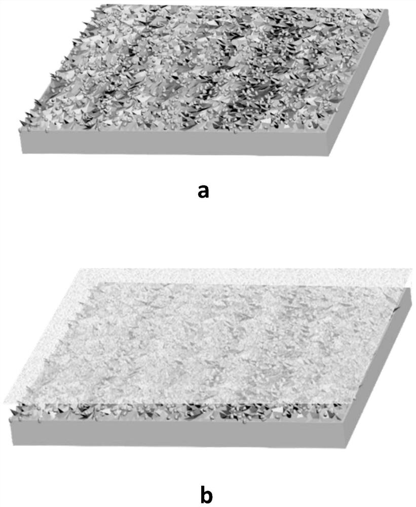
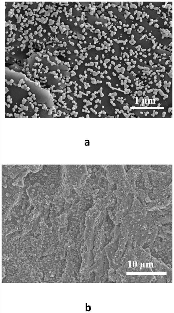
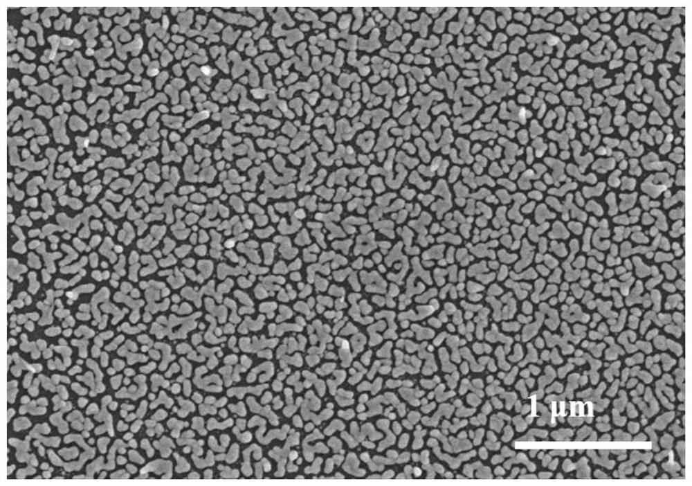
[0043] The schematic diagram of the base material is as figure 1 Shown in a and b, the higher the concentrations of hydroxylamine hydrochloride and tetrachloroauric acid, the larger the growth of gold particles, observing the scanning electron microscope images of Examples 1-3, Figure 5 a is the scanning electron microscope picture of embodiment 1, Figure 5 B is the scanning electron microscope figure of embodiment 2, Figure 5 c is the scanning electron microscope picture of embodimen...


PUM

PropertyMeasurementUnit
particle diameteraaaaaaaaaa
thicknessaaaaaaaaaa
particle diameteraaaaaaaaaa

Abstract

The invention discloses a surface-enhanced Raman scattering base material for gas detection and a preparation method thereof. The base material includes a base layer, the base layer is an inorganic material base provided with a rough working surface, and the working surface is loaded with in-situ grown gold Nano-island structure, the present invention forms a unique metal nano-island structure by growing metal nanoparticles in situ on an inorganic substrate with a rough surface, by regulating the size of the metal nanoparticles to 30-250nm, and the spacing is arranged at 1-80nm , the metal nanoparticles interact with each other, which significantly enhances the local electromagnetic field intensity on the surface of the metal nanoparticles and effectively improves the Raman signal intensity of the object to be detected. In addition, the combination of the substrate material and the metal-organic framework film material further increases the material The specific surface area can effectively increase the gas adsorption capacity, so that the surface-enhanced Raman scattering substrate can be applied to the detection of markers in the exhaled gas of cancer patients.

Description

technical field [0001] The invention relates to the technical field of surface-enhanced Raman scattering, in particular to a surface-enhanced Raman scattering base material for gas detection and a preparation method thereof. Background technique [0002] Volatile organic compounds (VOCs) in exhaled breath are products of human metabolism. For cancer patients, the production process of tumor cells is accompanied by changes in specific genes and proteins, and at the same time, peroxidation reactions occur on the surface of cell membranes, resulting in changes in VOCs in metabolites. On the other hand, cancer will affect the blood circulation of the human body, and the VOCs content in the exhaled air will also change accordingly through the exchange of the lungs. Not only the composition of VOCs in the breath of cancer patients is significantly different from that of healthy people, but also the content and proportion of VOCs are different for different cancers. Therefore, the...

Claims


Application Information

Patent Timeline
Patent Type & Authority Patents(China)
IPC IPC(8): G01N21/65
CPCG01N21/658
Inventor 王毅黄礼平张庆文李康蔡宇
Owner WENZHOU INST UNIV OF CHINESE ACAD OF SCI
Why Eureka
Try Eureka