Visual drainage tube

A drainage tube and inner tube technology, which is applied in the field of minimally invasive medical devices, can solve the problems such as the inability of the drainage tube to be used with the endoscope, the inability to accurately locate the position of the hematoma, and the difficulty in grasping the drainage process, so as to achieve convenient operation, improve drainage efficiency, damage reduction effect

Pending Publication Date: 2020-04-14
上海伴诚医疗科技有限公司
View PDF0 Cites 2 Cited by
  • Summary
  • Abstract
  • Description
  • Claims
  • Application Information

AI Technical Summary

Problems solved by technology

[0004] The technical problem to be solved by the present invention is that the existing drainage tube cannot be used with the endoscope, and the position of the hematoma cannot be precisely located. According to experience, repeated insertion of the drainage tube will cause harm to the patient, and it is difficult to grasp the drainage process.

Method used


Image

  • Visual drainage tube
  • Visual drainage tube
  • Visual drainage tube

Examples


Embodiment

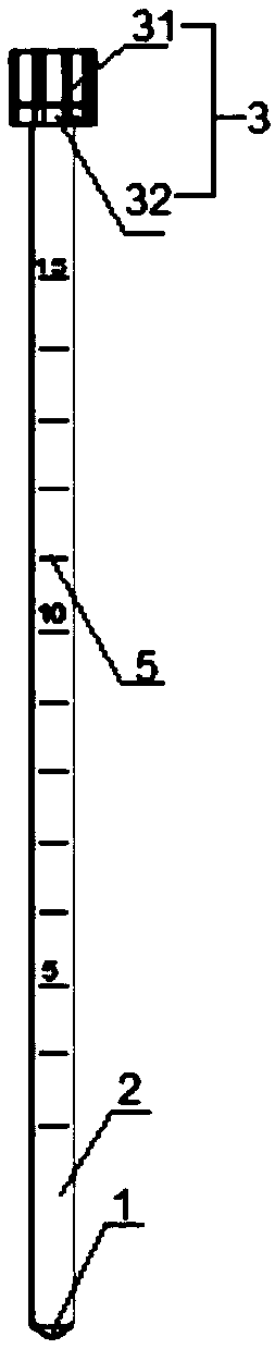
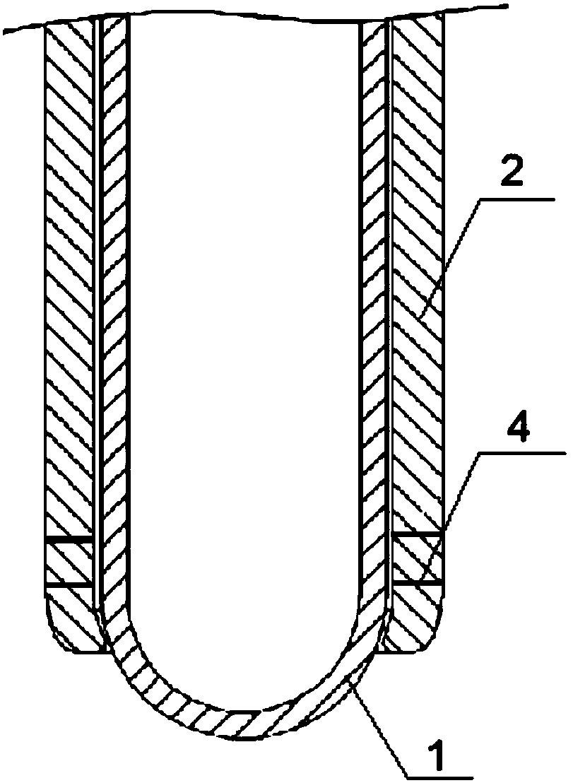
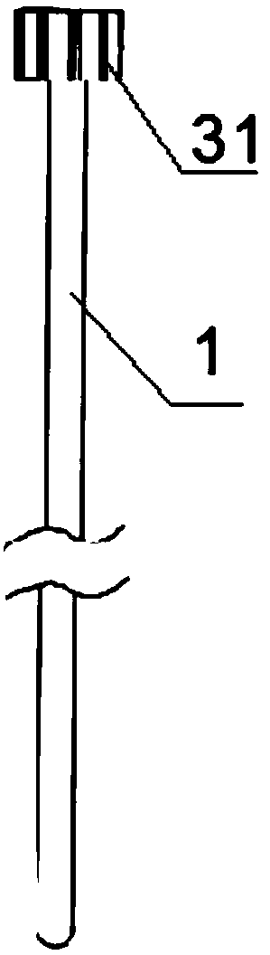
[0027] Embodiment: a kind of visual drainage tube, as figure 1 and figure 2 As shown, it includes an inner tube 1 for the endoscope to move therein and an outer tube 2 sleeved outside the inner tube 1 for the inner tube and the endoscope to move therein, and the inner tube 1 and the outer tube 2 are clearance fit; The tube 1 and the outer tube 2 are transparent and visible, and the thickness of the tube wall is uniform. One end of the inner tube 1 is closed, and the other end is provided with an opening for the endoscope to enter; both ends of the outer tube 2 are opened, and the inner tube 1 can One end of the outer tube 2 extends into the outer tube 2, and the closed end of the outer tube 2 close to the inner tube 1 extends toward the inner tube 1 in an O-shape and closely fits with the outer wall of the inner tube 1. Since the end of the outer tube 2 is open, the inner tube 1 The end part of the closed end protrudes from the outer tube 2, and the end of the outer tube 2 c...


PUM


Abstract

The invention provides a visual drainage tube, which relates to the field of minimally invasive medical instruments, and comprises an inner tube which is characterized in that an endoscope can move inthe inner tube, and an outer tube, wherein the inner tube is sleeved with the outer tube, the inner tube and the endoscope can move in the outer tube; the inner tube is in clearance fit with the outer tube; the inner tube and the outer tube are transparent and visible, and the tube walls are uniform in thickness; one end of the inner tube is closed, and the other end of the inner tube is providedwith an opening for the endoscope to enter; openings are formed in the two ends of the outer pipe; the end, close to the closed end of the inner pipe, of the outer pipe extends towards the inner pipeand is tightly attached to the outer wall of the inner pipe, and the end, close to the opening of the inner pipe, of the outer pipe is detachably connected with the inner pipe. The visual drainage tube has the advantages that the visual drainage tube is used in cooperation with the endoscope, the hematoma position can be accurately positioned, drainage is conducted at the same time, injuries caused by multiple times of insertion are reduced, meanwhile, the drainage situation can be observed in time, and the drainage effect is improved.

Description

technical field [0001] The invention relates to the field of minimally invasive medical devices, in particular to a visible drainage tube. Background technique [0002] The existing puncture and drainage technology for cerebral hematoma is mainly based on CT images before operation to perform in vitro positioning, and then puncture and drain according to the positioning point. However, during the puncture and drainage process, since the intracranial puncture process cannot be seen, it is performed entirely by the doctor's medical knowledge and experience, resulting in high technical requirements for the doctor's skills, resulting in the inability of the drainage tube to accurately puncture to the center of the hematoma. Difficult to master, resulting in poor hematoma drainage effect, slow recovery of patients, and even poor prognosis. [0003] Therefore, there is a need for a visual drainage tube that can be used with an endoscope, and the visual drainage tube can be used i...

Claims


Application Information

Patent Timeline
Patent Type & Authority Applications(China)
IPC IPC(8): A61M25/00
CPCA61M25/0023A61M25/0043A61M2025/0004
Inventor 兰津刘奇盛雨峰马捷
Owner 上海伴诚医疗科技有限公司
Why Eureka
Try Eureka