
System and method for color deconvolution of a slide image to assist in the analysis of tissue specimen

A deconvolution and component image technology, applied in the field of color deconvolution, can solve problems such as the nonlinearity of the color deconvolution equation

Active Publication Date: 2020-05-01
VENTANA MEDICAL SYST INC
View PDF5 Cites 0 Cited by
  • Summary
  • Abstract
  • Description
  • Claims
  • Application Information

AI Technical Summary

Problems solved by technology

[0015] However, in the case where both the reference dye vector and the dye component images are unknown and both need to be estimated, the color deconvolution equation is nonlinear

Method used


Image

  • System and method for color deconvolution of a slide image to assist in the analysis of tissue specimen
  • System and method for color deconvolution of a slide image to assist in the analysis of tissue specimen
  • System and method for color deconvolution of a slide image to assist in the analysis of tissue specimen

Examples


Embodiment Construction

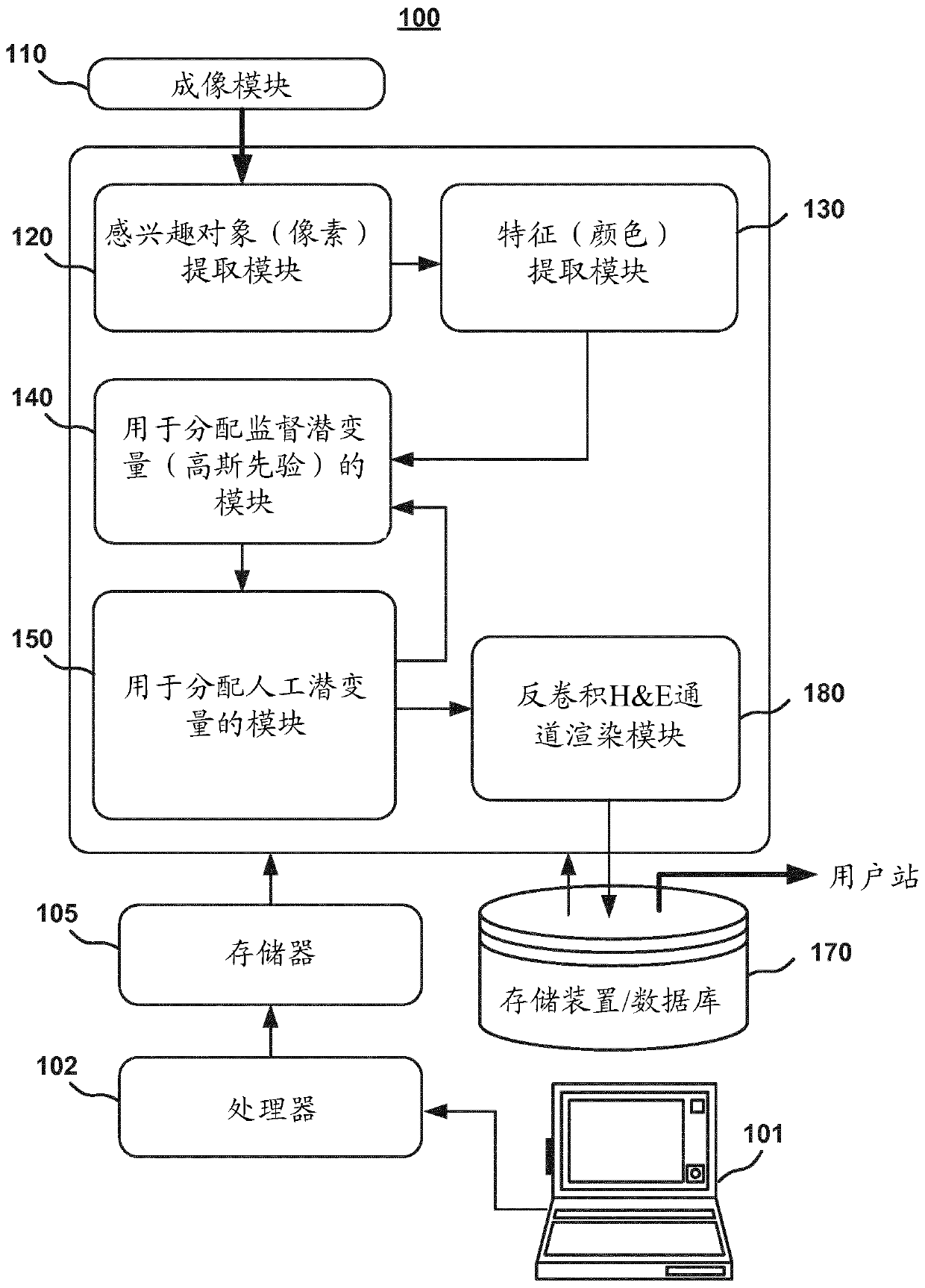
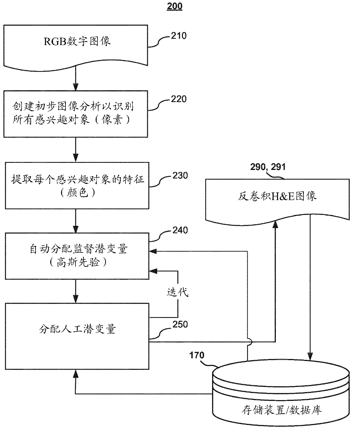
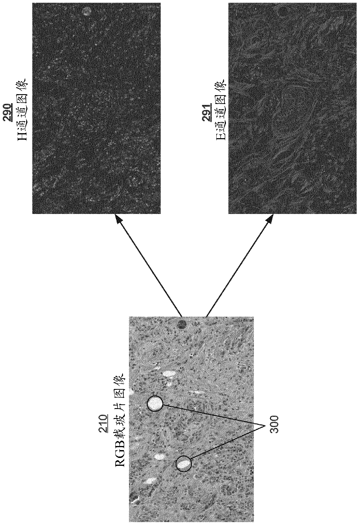
[0029] figure 1 Depicted is an automated computer-based analysis system 100 for converting three-channel stained slide digital RGB tissue images 210 ( image 3 ) is deconvolved into a two-channel component image, such as an H channel image 290 and an E channel image 291 ( image 3). The analysis system 100 may be adapted to analyze a biological sample, such as tissue provided on a glass slide. As used herein, the term "tissue sample" includes any type of biological sample, such as tissue sections, blood, cell cultures, and similar biological samples, which may be mounted on a glass slide.

[0030] The tissue image may be, for example, a multi-channel color image, such as RGB (or any equivalent color space, such as CYMK, Lab, YUV HSV, etc.) or other multi-channel color image of a tissue sample. In particular, the image data may comprise a pixel matrix consisting of a plurality of pixels representing an image of the tissue. A multi-channel color image may comprise a pluralit...


PUM


Abstract

A tissue analysis system and method for the spectral deconvolution of a RGB digital image obtained from a stained biological tissue sample, by estimating the stain component images that are obtained from a staining system configuration, where the reference stain vectors are assumed to be sampled from a known color distribution. The prior knowledge of stain variability of the staining system is adopted as initial reference stain vectors and statistical distribution of their variability. Based on the initial reference stain vectors distribution, the tissue analysis system determines both the reference stain vectors and stain component images of the input image. The image is then deconvoluted based on the reference stain vectors and stain component images.

Description

technical field [0001] The present disclosure generally relates to imaging systems and methods in digital pathology for cancer tissue diagnosis. More specifically, the present disclosure relates to color deconvolution of H&E-stained slide images that are digitally captured as multi-color channel images, such as red, green, and blue (RGB) channel images, for simultaneous Estimate dye component images and identify physiologically plausible reference dye vectors for assays. Background technique [0002] In the analysis of biological samples such as tissue sections, blood, cell cultures, etc., the biological sample is mounted on a slide and stained with one or more combinations of dyes and biomarkers, and the resulting tissue slide is subjected to Observe or image for further analysis. A tissue slide can include a biological sample, such as a tissue section from a human subject, that has been treated with a dye comprising a fluorophore or chromogen that binds to a protein, pro...

Claims


Application Information

Patent Timeline
Patent Type & Authority Applications(China)
IPC IPC(8): G06T7/90G06T5/00G01N21/00
CPCG06T2207/10024G06T2207/10056G06T2207/20076G06T2207/30024G06T7/90G06T5/73G06T7/0012G06V20/69
Inventor S·楚卡蓝舟
Owner VENTANA MEDICAL SYST INC
Learn More