

Unsupervised self-adaptive mammary gland lesion segmentation method
An adaptive model and image segmentation technology, applied in the field of image processing, can solve the problem of poor generalization ability of the segmentation model
- Summary
- Abstract
- Description
- Claims
- Application Information
AI Technical Summary
Problems solved by technology
Method used
Image

Examples
Embodiment 1
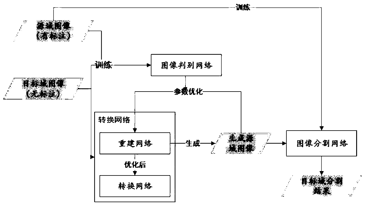
[0086] Such as figure 1 As shown, the present invention proposes a method for adaptive image conversion between different fields. Based on this method, even if there is a difference between the new data set and the labeled data set, there is no need to label the images in the new data set, but through image conversion Adaptive learning is performed on two datasets. As a result, the unlabeled dataset retains its high-level semantic information through adaptive transformation, and its shallow representations such as image style, texture, and brightness are converted into the features of the labeled dataset, so that the trained dataset in the labeled dataset can be directly The network model is directly applied to the new dataset.
[0087] In the adaptive image conversion method according to one embodiment of the present invention, comprise the following steps:
[0088] First, a dataset containing annotations is required as the source domain image, which can be regarded as an a...
Embodiment 2
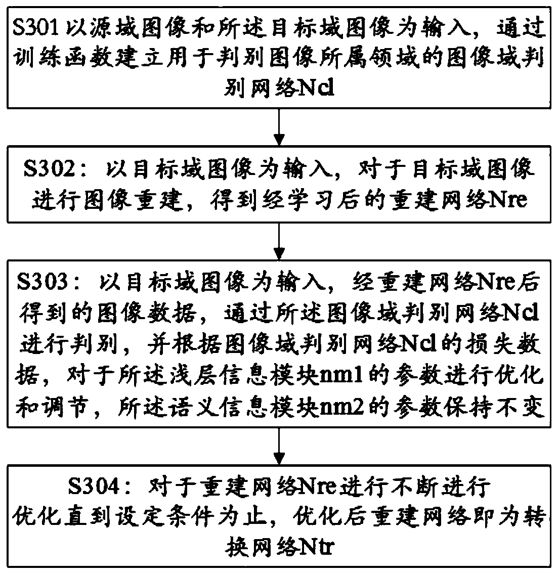
[0105] Such as figure 2 As shown, another embodiment of the present invention is an image segmentation method for unsupervised field adaptation, which can be specifically applied to computer-aided recognition of MRI images of breast lesions. It is mainly divided into: establishing an image domain discrimination network (S401), performing image reconstruction learning of target domain images (S402), optimizing based on the reconstruction network to obtain a conversion network (S403, S404), performing image processing on labeled source domain images. Segmentation network training (S405), converting the image of the target domain through the conversion network (S406), and then performing image segmentation on the converted target image by using the image segmentation network (S407).
[0106] In the above steps, the steps in S401, S402, S403, and S404 are the same as the corresponding steps in the first embodiment, and will not be repeated here.
[0107] Segmenting an annotated ...
Embodiment 3
[0116] see Figure 4 , the present embodiment provides a breast cancer screening device based on adaptive image segmentation, including:
[0117] An acquisition unit, configured to acquire source domain images in a source image set, where the images in the source domain image set contain marked feature regions, the source domain image set is a marked breast MRI image, and the feature regions are Lumps or areas of cancerous tissue to be marked;
[0118] It is also used to acquire a target domain image in the target domain image set, the target domain image is an unmarked breast MRI image, and the target domain image may contain an image part corresponding to a tumor or a cancerous tissue area;
[0119] An image domain discrimination unit, configured to take the source domain image and the target domain image acquired by the acquisition unit as input, and establish an image domain discrimination network Ncl for discriminating the domain to which the image belongs through a trai...
PUM
 Abstract
Description
Claims
Application Information