Medical ultrasonic image computer-aided diagnosis method

A computer-aided, ultrasonic image technology, applied in the directions of sonic diagnosis, ultrasonic/sonic/infrasonic diagnosis, infrasonic diagnosis, etc., can solve the problems of inconsistent conclusions, deviations from the actual situation, inability to effectively improve the efficiency and accuracy of diagnosis, etc. The effect of improving accuracy, improving diagnostic efficiency and accuracy

Pending Publication Date: 2020-06-02
刘涛
View PDF23 Cites 7 Cited by
  • Summary
  • Abstract
  • Description
  • Claims
  • Application Information

AI Technical Summary

Problems solved by technology

It is possible that the conclusion drawn by the medical auxiliary diagnosis system is consistent with the actual situation of the patient, and the conclusion is consistent; there are also conclusions drawn b

Method used


Image

  • Medical ultrasonic image computer-aided diagnosis method
  • Medical ultrasonic image computer-aided diagnosis method
  • Medical ultrasonic image computer-aided diagnosis method

Examples


Embodiment Construction

[0048]Reference will now be made in detail to the exemplary embodiments, examples of which are illustrated in the accompanying drawings. When the following description refers to the accompanying drawings, the same numerals in different drawings refer to the same or similar elements unless otherwise indicated. The implementations described in the following exemplary embodiments do not represent all implementations consistent with this application. Rather, they are merely examples of apparatuses and methods consistent with aspects of the present application as recited in the appended claims.

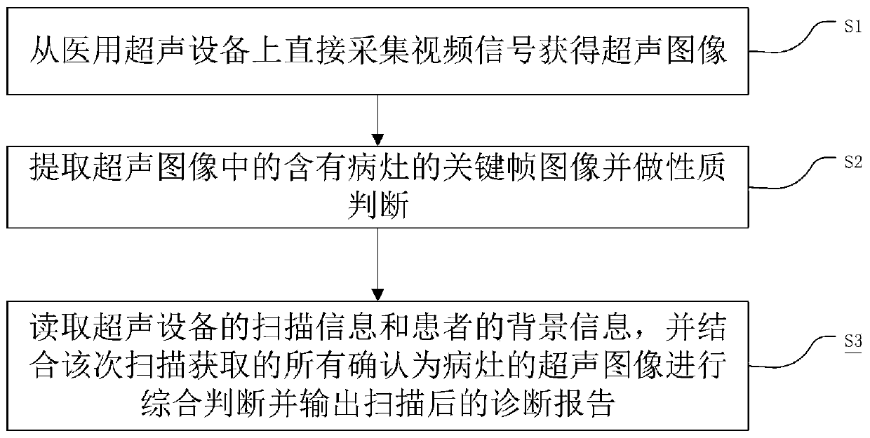
[0049] Such as figure 1 As shown, the present embodiment provides a method for computer-aided diagnosis of medical ultrasound images, including the following steps:

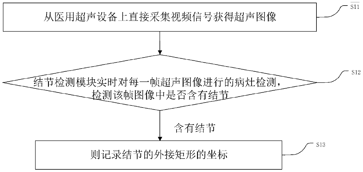
[0050] Step S1, directly collecting video signals from medical ultrasound equipment to obtain ultrasound images;

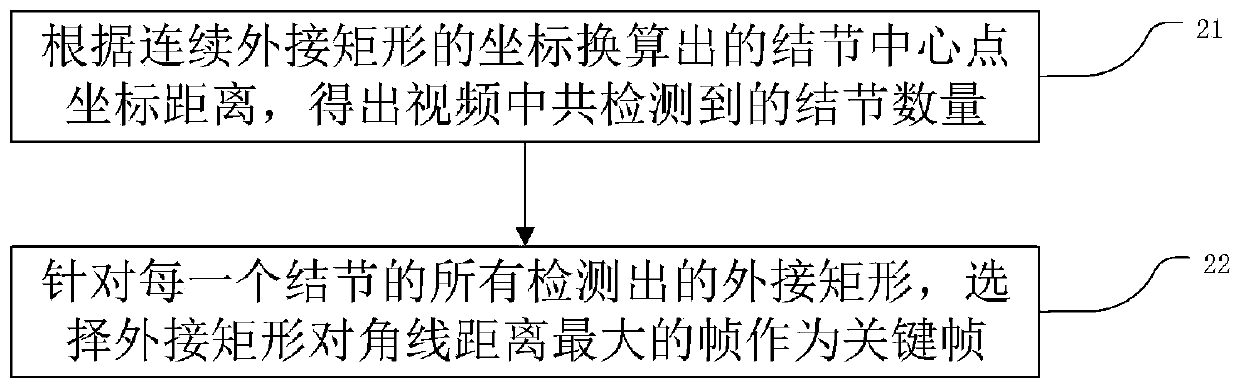
[0051] Step S2, extracting the key frame image containing the lesion in the ultrasound image and making a p...


PUM


Abstract

The invention relates to a medical ultrasonic image computer-aided diagnosis method. The method comprises the following steps: directly acquiring a video signal from medical ultrasonic equipment to obtain ultrasonic images; extracting key frame images containing a focus in the ultrasonic images and carrying out property judgment; performing comprehensive judgment in combination with all the ultrasonic images confirmed as the focus obtained by the scanning, outputting a scanned diagnosis report, and acquiring correction information and corresponding original focus images; re-training and updating a nodule key frame extraction module and a key frame nodule property judgment module according to the obtained correction information and the corresponding original focus images; and generating a new diagnosis report by using an updated model. The method can adapt to a current ultrasonic diagnosis scene, effectively assists a doctor in identifying a focus in the video frame images, forms a report and outputs the report, and improves the diagnosis efficiency and accuracy. The method can also acquire correction information given by a doctor, so that the system is continuously updated, and theaccuracy of auxiliary diagnosis can be continuously improved.

Description

technical field [0001] The present application relates to the technical field of ultrasonic image analysis, in particular to a method for computer-aided diagnosis of medical ultrasonic images. Background technique [0002] Ultrasonic examination (US examination) is to use the human body to observe the reflection of ultrasonic waves. Ultrasound examination, generally called US, is to irradiate the body with weak ultrasonic waves, and image the reflected waves of the tissues. In related technologies, ultrasonography has become an important and non-invasive detection method for displaying the structure and motion function of human organs. Medical ultrasound equipment is cheap and is used in hospitals and medical examination centers at all levels. Ultrasonography is cheap and has become an important means of early screening and diagnosis of many diseases. [0003] In the entire field of medical images, there are many technologies for computer-aided diagnosis, such as: CN10922...

Claims


Application Information

Patent Timeline
IPC IPC(8): A61B8/00
CPCA61B8/5215
Inventor 钱林学卜云芸庞浩刘涛
Owner 刘涛
Why Eureka
Try Eureka