Multi-modal imaging guided radiotherapy method, device and system

A multi-modal imaging and multi-modal image technology, which is applied in radiation therapy, X-ray/γ-ray/particle irradiation therapy, treatment, etc., can solve the problem that absorption imaging cannot meet the high-precision radiotherapy requirements, and improve radiotherapy Positioning accuracy, improve the effect of treatment effect

Pending Publication Date: 2020-06-23
SHANGHAI UNITED IMAGING HEALTHCARE
View PDF5 Cites 2 Cited by
  • Summary
  • Abstract
  • Description
  • Claims
  • Application Information

AI Technical Summary

Problems solved by technology

Therefore, a single absorption imaging cannot meet the high-precision radiotherapy needs

Method used


Image

  • Multi-modal imaging guided radiotherapy method, device and system
  • Multi-modal imaging guided radiotherapy method, device and system
  • Multi-modal imaging guided radiotherapy method, device and system

Examples


Embodiment 1

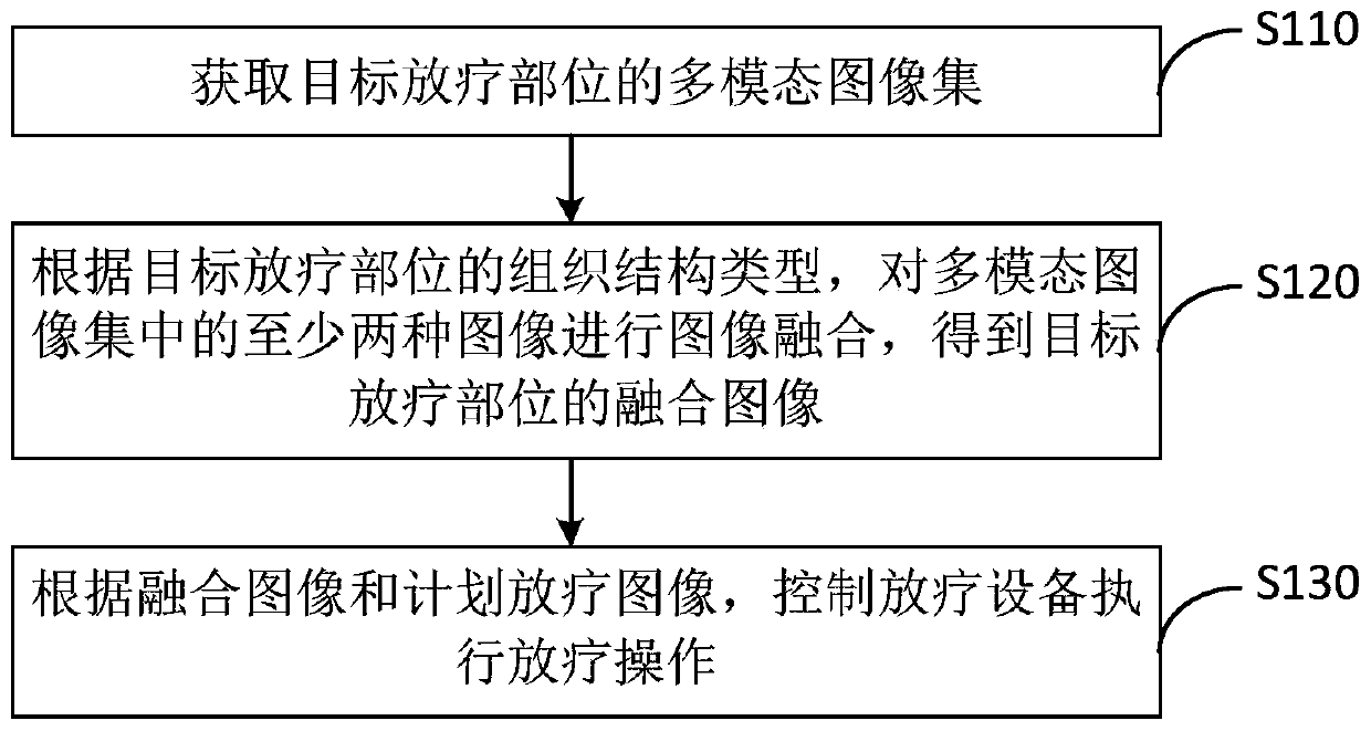
[0028] figure 1 It is a flowchart of a multi-modal imaging-guided radiotherapy method provided by the first embodiment of the present invention. This embodiment is applicable to the situation of image-guided precise radiotherapy. The method can be executed by a multi-modal imaging-guided radiotherapy device The device can be implemented in software and / or hardware, and the device can be configured in terminal equipment. It includes the following steps:

[0029] S110. Acquire a multi-modal image set of the target radiotherapy site, where the multi-modal image set includes at least two images among an absorption image, a phase image, and a dark field image.

[0030] In phase contrast imaging, each image recorded by the detector in the multimodal imaging device is a mixed information image containing the absorption, phase, and dark field of the object. The three contrast mechanisms correspond to the different composition and structure of the object. Therefore, it is necessary to extr...

Embodiment 2

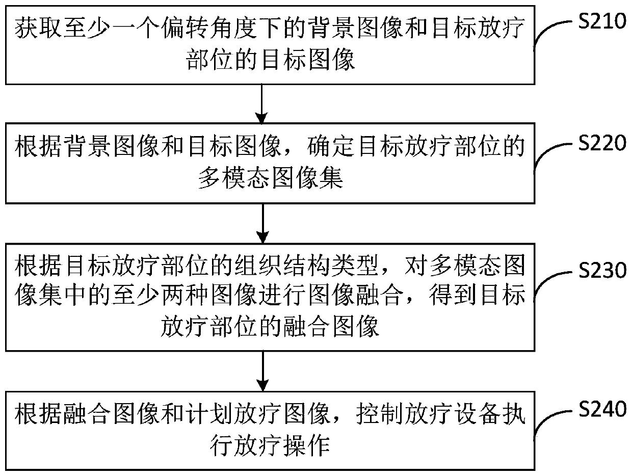
[0052] figure 2 It is a flowchart of a multi-modal imaging-guided radiotherapy method provided in the second embodiment of the present invention. The technical solution of this embodiment is a further refinement on the basis of the foregoing embodiment. Optionally, the acquiring a multi-modal image set of the target radiotherapy site includes: acquiring a background image at at least one deflection angle and a target image of the target radiotherapy site; determining the background image and the target image A collection of multi-modal images of the target radiotherapy site.

[0053] The specific implementation steps of the embodiment of the present invention include:

[0054] S210: Acquire a background image and a target image of a target radiotherapy site under at least one deflection angle.

[0055] Among them, the deflection angle refers to the angle between the X-ray emitted by the light source of the multi-modal imaging device and the plane of the target radiotherapy site. Am...

Embodiment 3

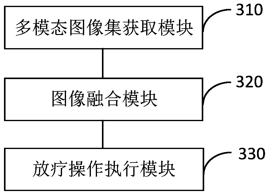
[0073] image 3 It is a schematic diagram of a multi-modal imaging-guided radiotherapy device provided in the third embodiment of the present invention. This embodiment is applicable to the situation of image-guided precise radiotherapy. The device can be implemented in software and / or hardware, and the device can be configured in a terminal device. The radiotherapy apparatus guided by multimodal imaging includes: a multimodal image set acquisition module 310, an image fusion module 320, and a radiotherapy operation execution module 330.

[0074] Wherein, the multi-modal image set acquisition module 310 is used to acquire a multi-modal image set of the target radiotherapy site, wherein the multi-modal image set includes at least two images of an absorption image, a phase image and a dark field image;

[0075] The image fusion module 320 is configured to perform image fusion on at least two images in the multimodal image set according to the tissue structure type of the target radio...


PUM


Abstract

The embodiment of the invention discloses a multi-modal imaging guided radiotherapy method, device and system. The method comprises the steps that a multi-modal image set of a target radiotherapy partis acquired, and the multi-modal image set comprises at least two images of an absorption image, a phase image and a dark field image; according to the tissue structure type of the target radiotherapy part, performing image fusion on at least two images in the multi-modal image set to obtain a fused image of the target radiotherapy part; and controlling radiotherapy equipment to execute radiotherapy operation according to the fused image and the planned radiotherapy image. According to the embodiment of the invention, the method achieves the fusion of the at least two types of images of the target radiotherapy part, solves a problem that the single imaging cannot meet the high-precision radiotherapy demands, improves the radiotherapy positioning precision, enables the radiotherapy equipment to carry out the precise radiotherapy of the target radiotherapy part, and improves the treatment effect of radiotherapy.

Description

Technical field [0001] The embodiments of the present invention relate to the technical field of radiotherapy, and in particular to a multimodal imaging guided radiotherapy method, device and system. Background technique [0002] Radiotherapy is one of the important treatment methods for tumor treatment. Precision radiotherapy for tumors is the application of various technologies or equipment to make the target area highly conformable to ensure that the prescribed dose in the treatment is concentrated in the tumor area to the maximum extent and the surrounding normal organs Get maximum protection. Therefore, it is particularly important to achieve precise positioning of the tumor area. [0003] Image-guided radiotherapy is based on three-dimensional conformal radiotherapy with the addition of time factors, and fully considers the movement of the target area and surrounding normal organs during the treatment process and the positioning error between the radiotherapy divisions. Befo...

Claims


Application Information

Patent Timeline
IPC IPC(8): G06T5/50A61N5/10
CPCG06T5/50A61N5/103A61N5/1048A61N5/1049G06T2207/20221A61N2005/1061
Inventor 韩华杰
Owner SHANGHAI UNITED IMAGING HEALTHCARE
Why Eureka
Try Eureka