Tumor and organ position ultrasonic image real-time monitoring system for radiotherapy

A real-time monitoring system and ultrasonic image technology, applied in the field of medical devices, can solve problems such as increasing the burden of human resources, affecting the coverage and fixation effect of external fixation body membranes, and interfering with the spatial direction of the radiotherapy machine head

Pending Publication Date: 2021-08-20
程明霞
View PDF0 Cites 0 Cited by
  • Summary
  • Abstract
  • Description
  • Claims
  • Application Information

AI Technical Summary

Problems solved by technology

These placement methods will affect the coverage and fixation effect of the external fixation membrane on the body; mechanical devices and robotic arms will interfere with the rotatable spatial direction of the radiotherapy machine head when the beam is emitted, and the method of binding and fixing the probe with tape or belt is also lacking in stability. Repeatability of sex and position
And in these placements, the probe interferes with the selection of the optimal field
It is even more impossible to place multiple ultrasound probes at the same time for multi-level and multi-angle real-time monitoring
[0007] Secondly, the interpretation of ultrasound images is highly dependent on the operator. The identification of anatomical organs and their signs in ultrasound images in real-time monitoring usually requires the presence of professional ultrasound doctors, which will increase the burden of human resources and is almost impossible to be widely used in conventional radiotherapy. popularization of
If non-ultrasound personnel randomly select the position of the ultrasonic probe and monitor by feeling, it is impossible to achieve the purpose of real-time monitoring of precise radiotherapy

Method used


Image

  • Tumor and organ position ultrasonic image real-time monitoring system for radiotherapy
  • Tumor and organ position ultrasonic image real-time monitoring system for radiotherapy
  • Tumor and organ position ultrasonic image real-time monitoring system for radiotherapy

Examples


Deformed example 2

[0084] In the second modification, the same symbols are used for the same structures as those in the embodiment, and corresponding descriptions are omitted.

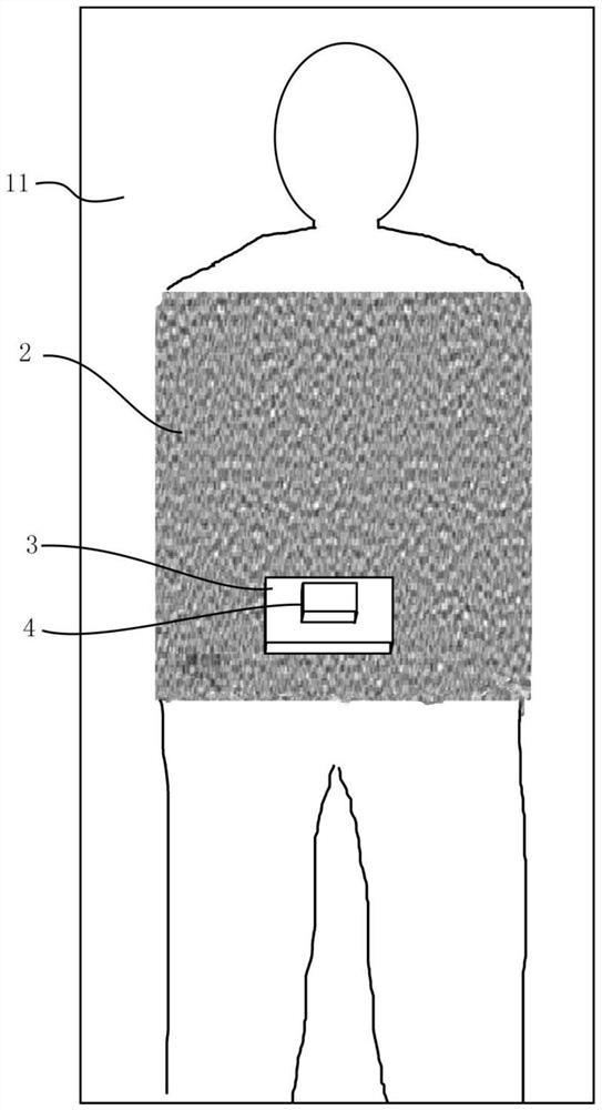
[0085] In the above-mentioned embodiment, the fixed body film 2 and the fixed part 3 are printed parts integrally formed by 3D printing. In the second modification, the fixed part 3' can also use standard parts of different sizes and models produced by traditional methods. .

[0086] In this modified example, the contact surface of the fixed part 3' and the fixed body film 2' is provided with a hook surface, and when the fixed body film 2' is 3D printed, a rough surface is printed at the position where the fixed part 3' is installed . The above-mentioned rough surface and the hook surface form a mounting assembly for installing the fixed part on the fixed body membrane, so that the fixed part 3' can be fixed on the fixed body membrane 2 in the form of Velcro, and further to the ultrasonic probe 4 to fix.

[0087] The ...


PUM


Abstract

The invention provides a tumor and organ position ultrasonic image real-time monitoring system for radiotherapy. The system is characterized by comprising radiotherapy device, a fixing body membrane, a fixing part, an ultrasonic probe and a processing device, wherein the radiotherapy device comprises a treatment bed and a radiotherapy irradiation device, and the radiotherapy irradiation device is used for irradiating a patient according to a preset irradiation range; the fixing body membrane is matched with the treatment bed to fix the patient; the fixing part is arranged on the body membrane and located outside the irradiation range; the ultrasonic probe is arranged on the fixing part and attached to the body surface of the patient; and the fixing body membrane is provided with an opening part matched with the fixing position of the ultrasonic probe, the processing device is provided with an ultrasonic image recognition part and a radiotherapy control part, the ultrasonic image recognition part recognizes an ultrasonic image according to a marker pre-calibrated by a radiotherapy doctor so as to recognize the real-time position of a tumor or an organ or an implant or the like of the patient, and the radiotherapy control part controls the radiotherapy irradiation device to emit beams for irradiation according to the irradiation range when the real-time position of the marker is within a preset threshold range.

Description

technical field [0001] The invention belongs to the field of medical devices, and in particular relates to a real-time monitoring system for ultrasonic images of tumor and viscera positions used for radiotherapy. Background technique [0002] Radiation therapy (radiotherapy) is one of the main treatments for cancer. Radiation therapy uses ionizing radiation such as high-energy X-rays, electrons, and proton heavy ions to kill tumor tissues in the body. Because of the penetrability of radiation, high-energy radiation will inevitably damage the normal organs and tissues of the human body while killing tumors. The damage varies according to the irradiation site and the irradiation dose, such as skin damage, radiation pneumonitis ( Chest tumors such as lung cancer), radiation enteritis (abdominal tumors), impaired swallowing function (head and neck tumors), etc. Severe injuries can lead to bowel perforation, blindness, paralysis, and even death. Therefore, radiotherapy uses a ...

Claims


Application Information

Patent Timeline
Patent Type & Authority Applications(China)
IPC IPC(8): A61N5/10
CPCA61N5/1049A61N2005/1058A61N2005/1055A61N2005/1062
Inventor 程明霞
Owner 程明霞
Why Eureka
Try Eureka