Radiotherapy in-vivo dose monitoring method

A radiation therapy and dose technology, applied in radiation therapy, therapy, X-ray/γ-ray/particle irradiation therapy, etc., can solve problems such as position deviation, unpredictable skin changes, and unreliable patient dose monitoring results

Pending Publication Date: 2021-05-28
THE SECOND AFFILIATED HOSPITAL ARMY MEDICAL UNIV
View PDF4 Cites 2 Cited by
  • Summary
  • Abstract
  • Description
  • Claims
  • Application Information

AI Technical Summary

Problems solved by technology

[0005] However, radiotherapy is a long-term phase, and it goes through a long period of time, and the changes of the human skin during this period are unpredictable
At the same time, because the positioning line is marked on the skin or thermoplastic film instead of the target area (the target area is in the body), the human body is composed of a large number of soft tissue structures such as non-rigid fat and muscle, and there may be a certain displacement between various organs, and there may be The positioning line has been aligned with the body surface, but the position of the target area is deviated. In this case, the patient’s in-body dose monitoring results are not reliable; Exposure to normal tissue outside the body may cause additional harm to the patient

Method used


Image

  • Radiotherapy in-vivo dose monitoring method

Examples


Embodiment 1

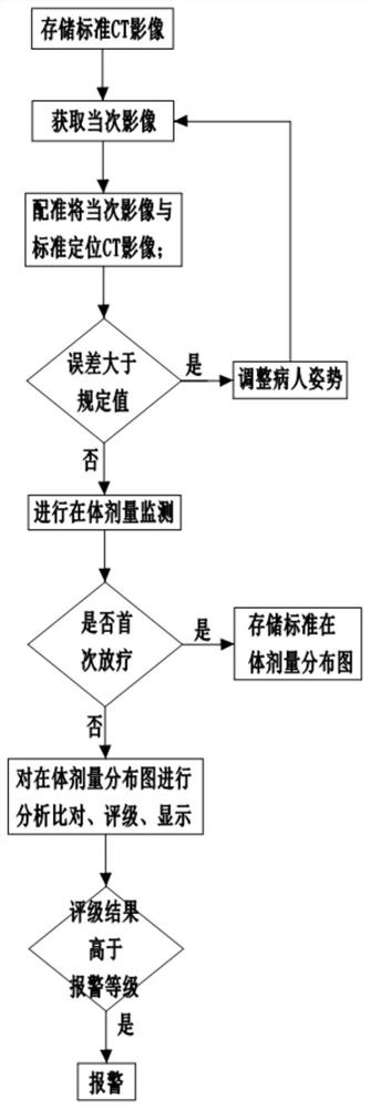
[0042]It should be noted that the implementation of this application is based on an in-body dose monitoring system for radiotherapy, which includes scanning equipment, radiotherapy equipment, a processor, and a medical database. The medical database stores the patient's disease type, treatment plan, planned in-body dose and corresponding positioning CT images.

[0043] Medical equipment includes CT image-guided scanning equipment, gantry and radiation therapy equipment. The radiotherapy equipment includes a treatment machine head, an image receiver and a treatment couch. The treatment machine head is installed on a frame, the treatment couch is located under the treatment machine head, and the treatment couch is located between the ray transmitter and the image receiver. In this embodiment, the image receiver is EPID.

[0044] The scanning device is a two-dimensional imaging device or a three-dimensional imaging device. In the present embodiment, the scanning device is a CBCT...

Embodiment 2

[0066] Anesthesia is usually not used during radiation therapy because most of the radiation therapy is painless for the patient. During the course of radiotherapy, the patient may move slightly due to numbness, itching, etc. If radiotherapy continues, the site of radiotherapy will be inaccurate. However, due to the small movement, if the radiotherapy is stopped, the patient's It is not only inefficient to continue the treatment after the position is adjusted, but also the operation is complicated, and the continuous treatment time of the patient's single treatment cannot be guaranteed.

[0067] Therefore, in the present embodiment, the treatment bed includes a first bed board, a lifting part, a second bed board and a rotating part from top to bottom; the first bed board and the second bed board have the same cross section, and the lifting part includes four electric telescopic rods, The bases of the four electric telescopic rods are respectively fixed to the four corners of t...


PUM


Abstract

The invention belongs to the technical field of dose monitoring, and particularly provides a radiotherapy in-vivo dose monitoring method, which comprises the following steps: S0, acquiring a CT image of a body part, needing to be irradiated, of a patient, making a radiotherapy plan according to the CT image, obtaining a positioning CT image of a target region, and storing the positioning CT image of the target region as a standard CT image; S1, acquiring an image of an in-vivo anatomical structure of a current treatment part of a patient through scanning equipment, and recording the image as a current image; S2, registering the current image with a standard positioning CT image; if the registration result is that the error is greater than the specified value, adjusting the posture of the patient, and returning to the S1; if the registration result accords with a specified value, entering S3; and S3, carrying out radiotherapy on the patient by using radiotherapy equipment, and monitoring the in-vivo dose of the patient. Compared with the prior art, the method has the advantages that additional injury to the patient can be avoided as much as possible, the treatment effect is better, and effectiveness and accuracy of in-vivo dose monitoring can be guaranteed.

Description

technical field [0001] The invention belongs to the technical field of dose monitoring, and in particular relates to a method for monitoring in vivo dose of radiotherapy. Background technique [0002] Radiotherapy is one of the important treatment methods for cancer at present. When precise radiotherapy techniques such as intensity-modulated radiotherapy and three-dimensional stereotactic radiotherapy are used to treat patients, in order to ensure the therapeutic effect, it is necessary to ensure that the radiotherapy target area receives the correct radiation dose. In the case of ensuring the accurate beam dose of the treatment machine, the deviation of the irradiation position is one of the important factors affecting the patient's exposure dose. [0003] The current common process is to obtain the patient's CT positioning image based on the results of CT simulation positioning, so as to understand the spatial image of the patient's internal anatomical position and the pos...

Claims


Application Information

Patent Timeline
Patent Type & Authority Applications(China)
IPC IPC(8): A61N5/10
CPCA61N5/1001A61N5/103A61N5/1039A61N5/1049A61N5/1071A61N2005/1061A61N2005/1092
Inventor 周一兵孙建国赵利荣段小娟周军
Owner THE SECOND AFFILIATED HOSPITAL ARMY MEDICAL UNIV
Why Eureka
Try Eureka