
Medical image processing method and device, equipment and storage medium

A medical image and processing method technology, applied in the field of image processing, can solve the problem of low disease recognition accuracy, and achieve the effect of improving the effect

Pending Publication Date: 2020-06-26
BOE TECH GRP CO LTD
View PDF4 Cites 5 Cited by
  • Summary
  • Abstract
  • Description
  • Claims
  • Application Information

AI Technical Summary

Problems solved by technology

[0003] In view of the shortcomings of the existing methods, this application proposes a medical image processing method, device, equipment and storage medium to solve the technical problems of low accuracy in image detection and disease identification existing in the prior art

Method used


Image

  • Medical image processing method and device, equipment and storage medium
  • Medical image processing method and device, equipment and storage medium
  • Medical image processing method and device, equipment and storage medium

Examples


Embodiment Construction

[0027] The present application is described in detail below, and examples of embodiments of the present application are shown in the drawings, wherein the same or similar reference numerals denote the same or similar components or components having the same or similar functions throughout. Also, detailed descriptions of known technologies will be omitted if they are not necessary to illustrate the features of the present application. The embodiments described below by referring to the figures are exemplary only for explaining the present application, and are not construed as limiting the present application.

[0028] Those skilled in the art can understand that, unless otherwise defined, all terms (including technical terms and scientific terms) used herein have the same meanings as commonly understood by those of ordinary skill in the art to which this application belongs. It should also be understood that terms, such as those defined in commonly used dictionaries, should be ...


PUM


Abstract

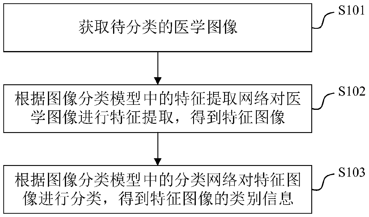
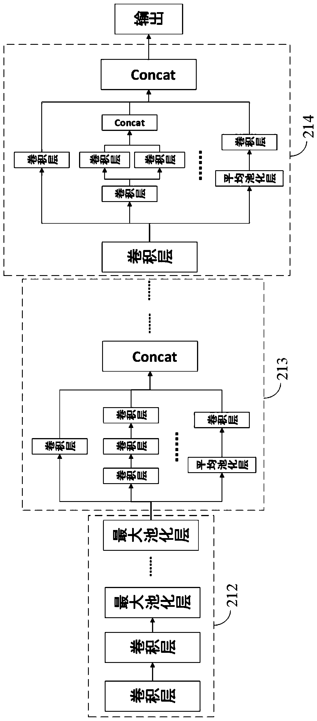
The invention provides a medical image processing method and device, equipment and a storage medium. The medical image processing method comprises the steps of obtaining a to-be-classified medical image; performing feature extraction on the medical image according to a feature extraction network in the image classification model to obtain a feature image; and classifying the feature images according to a classification network in the image classification model to obtain category information of the feature images. According to the embodiment of the invention, the method can achieve the more accurate prediction of the types of to-be-classified medical images, and the prediction result can be used for assisting a doctor in the accurate and quick diagnosis of a disease of a patient, thereby improving the auxiliary diagnosis effect.

Description

technical field [0001] The present application relates to the technical field of image processing, and in particular, the present application relates to a medical image processing method, device, device, and storage medium. Background technique [0002] Doctors usually need to use medical images (X-ray images, ultrasound images, etc.) as a reference for disease diagnosis when performing diagnosis and treatment. At present, doctors' manual diagnosis is mainly used, and the diagnosis efficiency is low. There are some auxiliary diagnoses based on medical images, but they usually give specific indicators or perform rough detection and disease identification on medical images. The detection accuracy is not high and the execution efficiency is low, and the auxiliary effect on diagnosis is poor. Contents of the invention [0003] Aiming at the shortcomings of the existing methods, the present application proposes a medical image processing method, device, equipment and storage me...

Claims


Application Information

Patent Timeline
Patent Type & Authority Applications(China)
IPC IPC(8): G06K9/62G06K9/46G16H30/20G16H50/20
CPCG16H30/20G16H50/20G06V10/40G06F18/241
Inventor 欧歌吴琼杨志明马小惠唐大伟
Owner BOE TECH GRP CO LTD
Learn More