
Fundus retina blood vessel recognition and quantification method, device and equipment and storage medium

A retinal blood vessel and quantification method technology, applied in the field of devices, fundus retinal blood vessel identification and quantification methods, equipment and storage media, can solve the problems of low quantification accuracy, long time consumption, and great influence on arteriovenous vessel identification and classification, and achieve The effect of improving recognition accuracy and improving quantization accuracy

Pending Publication Date: 2020-06-26
PING AN TECH (SHENZHEN) CO LTD
View PDF0 Cites 25 Cited by
  • Summary
  • Abstract
  • Description
  • Claims
  • Application Information

AI Technical Summary

Problems solved by technology

Calculating the diameter ratio of retinal arteriovenous vessels requires accurate classification of arteriovenous vessels. Traditional fundus retinal diagnosis relies on doctors to observe fundus images and obtain diagnostic results based on their own medical experience. This method takes a long time and is The workload is heavy, and the identification and classification of arteriovenous vessels are greatly affected by subjectivity
With the development of computer image processing technology, at present, fundus color photos are used to extract fundus retinal blood vessels. Because the fundus color photos have the characteristics of uneven brightness, complex interlacing of blood vessels and background colors, and small difference between arteries and veins, it is very important for arteriovenous blood vessel identification and recognition. Classification causes certain difficulties, and the existing research on automatic identification of arteriovenous vessels mainly realizes local arteriovenous vessel identification based on color or partial structure of blood vessels, but the identification accuracy is still limited, which leads to low quantification accuracy

Method used

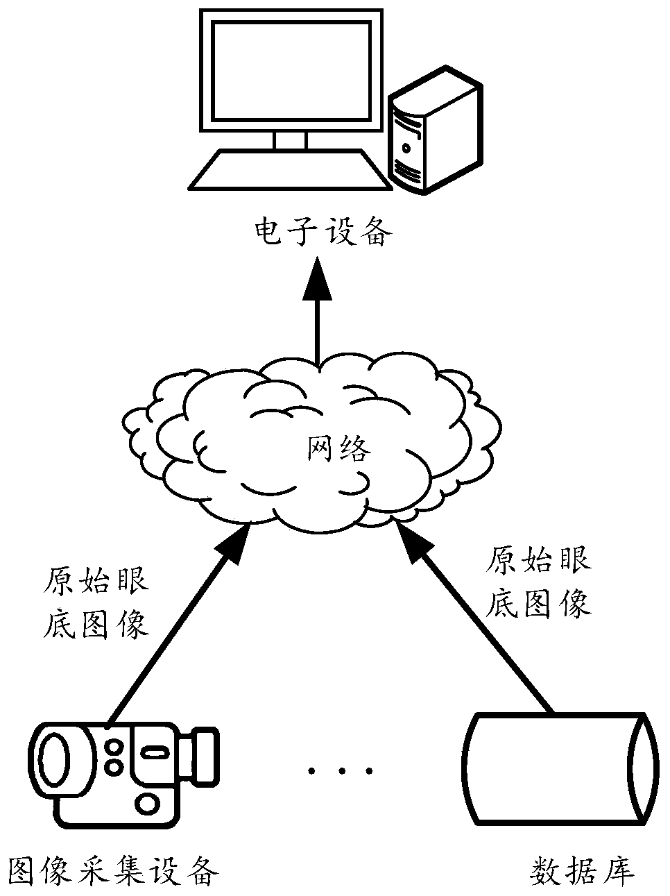

Image

  • Fundus retina blood vessel recognition and quantification method, device and equipment and storage medium
  • Fundus retina blood vessel recognition and quantification method, device and equipment and storage medium
  • Fundus retina blood vessel recognition and quantification method, device and equipment and storage medium

Examples


Embodiment Construction

[0064] In order to enable those skilled in the art to better understand the solution of the present application, the technical solution in the embodiment of the application will be clearly and completely described below in conjunction with the accompanying drawings in the embodiment of the application. Obviously, the described embodiment is only It is an embodiment of a part of the application, but not all of the embodiments. Based on the embodiments in this application, all other embodiments obtained by persons of ordinary skill in the art without creative efforts shall fall within the scope of protection of this application.

[0065] The terms "comprising" and "having" and any variations thereof appearing in the specification, claims and drawings of this application are intended to cover non-exclusive inclusion. For example, a process, method, system, product or device comprising a series of steps or units is not limited to the listed steps or units, but optionally also incl...


PUM


Abstract

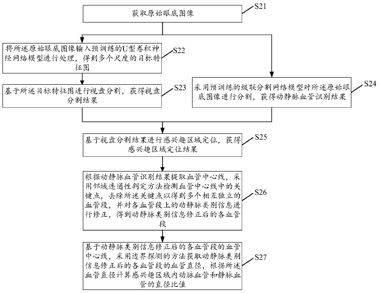
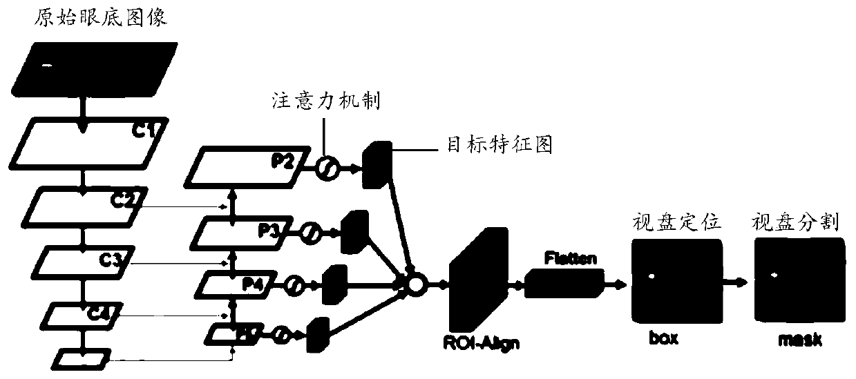
The invention provides a fundus retinal vessel recognition and quantification method, device and equipment and a storage medium, and the method comprises the steps: inputting an original fundus imageinto a pre-trained U-shaped convolutional neural network model for processing, and obtaining a target feature map; performing optic disk segmentation based on the target feature map; segmenting the original fundus image to obtain an arteriovenous blood vessel recognition result; carrying out region-of-interest positioning based on the optic disk segmentation result; extracting a blood vessel center line according to the arteriovenous blood vessel recognition result, detecting key points in the blood vessel center line, removing the key points to obtain a plurality of mutually independent bloodvessel sections, and correcting arteriovenous category information on each blood vessel section; and based on the extracted blood vessel center line, obtaining the blood vessel diameter of each bloodvessel section after category information correction, and then quantifying arteriovenous blood vessels in the region of interest. According to the embodiment of the invention, the fundus retina artery and vein vessel identification precision is improved, and the quantization precision is further improved.

Description

technical field [0001] The present application relates to the technical field of image processing, and in particular to a fundus retinal blood vessel identification and quantification method, device, equipment and storage medium. Background technique [0002] The fundus retinal arteries and veins have always been the focus of medical research, especially those within 1pd-1.5pd (Papillary Diameter, optic disc diameter) from the center of the optic disc. Evidence for early diagnosis of systemic and blood diseases, such as cardiovascular disease, diabetes, hypertension, etc. Calculating the diameter ratio of retinal arteriovenous vessels requires accurate classification of arteriovenous vessels. Traditional fundus retinal diagnosis relies on doctors to observe fundus images and obtain diagnostic results based on their own medical experience. This method takes a long time and is The workload is heavy, and the identification and classification of arteriovenous vessels are greatl...

Claims


Application Information

Patent Timeline
IPC IPC(8): G06T7/00G06T7/10G06T7/13G06T7/62A61B3/12G06N3/04
CPCG06T7/0012G06T7/10G06T7/13G06T7/62A61B3/12G06T2207/20104G06T2207/30101G06T2207/30041G06N3/045
Inventor 柳杨王瑞王立龙吕彬吕传峰
Owner PING AN TECH (SHENZHEN) CO LTD
Learn More