Chest radiography lung field segmentation model establishment based on multi-scale feature fusion and segmentation method

A multi-scale feature and segmentation model technology, applied in the field of medical image analysis, can solve problems such as inaccuracy and under-segmented edge segmentation of lung fields, and achieve the effect of improving segmentation accuracy and segmentation effect.

Active Publication Date: 2020-07-17
NORTHWEST UNIV
View PDF11 Cites 12 Cited by
  • Summary
  • Abstract
  • Description
  • Claims
  • Application Information

AI Technical Summary

Problems solved by technology

[0005] In order to solve the deficiencies in the prior art, the present invention provides a chest X-ray lung field segmentation model establishment and segmentation method based ...

Method used


Image

  • Chest radiography lung field segmentation model establishment based on multi-scale feature fusion and segmentation method
  • Chest radiography lung field segmentation model establishment based on multi-scale feature fusion and segmentation method
  • Chest radiography lung field segmentation model establishment based on multi-scale feature fusion and segmentation method

Examples


Embodiment Construction

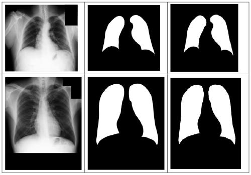
[0045] The data set used in the specific embodiment of the present invention is an X-ray chest image obtained from a hospital. The data set contains 138 cases of chest X-ray images. The data set is randomly divided into three parts, and the network is evaluated by three-fold cross-validation. Take the average of the three folds as the final result.

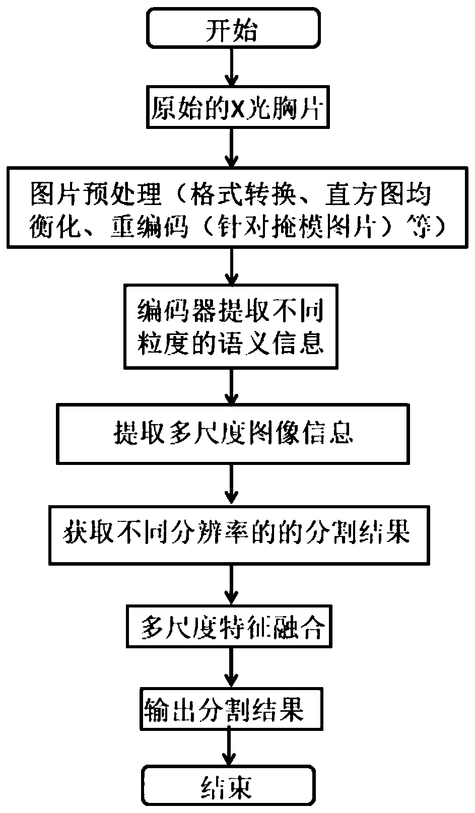
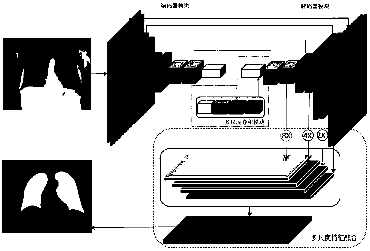
[0046] The method for establishing a chest X-ray lung field segmentation model based on multi-scale feature fusion disclosed in the specific embodiments of the present invention specifically includes the following steps:

[0047] Step 1, preprocessing of X-ray chest film;

[0048] Step 1.1, X-ray chest film may have low image contrast due to equipment or lighting, that is, the overall image is dark or bright. Performing histogram equalization on the image can improve the contrast of the image and reduce the gray scale. The value is mapped to 0-255, and saved as PNG or JPG format, and the preprocessed image is used as the input du...


PUM


Abstract

The invention discloses a chest radiography lung field segmentation model establishment based on multi-scale feature fusion and segmentation method. A segmentation model establishing method comprisesthe following steps: firstly, processing an X-ray chest radiograph; obtaining a preprocessed picture and a recoded mask picture, then constructing an X-ray chest radiography lung field segmentation network based on multi-scale convolution and a feature pyramid, and finally training the segmentation network by using the preprocessed picture as an input of the segmentation network and using the recoded mask picture as an output of the segmentation network to obtain a trained segmentation model; and based on the obtained segmentation model, preprocessing any to-be-processed X-ray chest radiograph, and inputting the preprocessed X-ray chest radiograph into the segmentation model to obtain a lung field segmentation result. According to the method, multi-resolution feature fusion is provided incombination with a feature pyramid theory, wherein the segmentation results with different resolutions can be fused, so that the segmentation effect is improved.

Description

technical field [0001] The invention belongs to the technical field of medical image analysis, and relates to a chest X-ray lung field segmentation model establishment and segmentation method based on multi-scale feature fusion. Background technique [0002] Chest X-ray (CXR) imaging is currently the most popular and available diagnostic tool for health monitoring and diagnosis of several lung diseases including pneumonia, tuberculosis, cancer, etc. But detecting these diseases from CXR is a very complex process and requires the involvement of radiologists. Moreover, millions of CXRs are generated every year. According to the National Health Service (UK), more than 22.9 million X-ray images were produced in the UK in 2017 / 18, accounting for all imaging equipment including magnetic resonance (MRI) and computed tomography (CT). 55.63%. The global shortage of radiologists suggests that this is a considerable diagnostic workload. [0003] An important step in the computer-ai...

Claims


Application Information

Patent Timeline
IPC IPC(8): G06T7/174G06T7/143G06T7/00G06T5/40G06K9/62G06N3/04G06N3/08
CPCG06T7/174G06T7/143G06T7/0012G06T5/40G06N3/08G06T2207/10116G06T2207/20016G06T2207/30061G06N3/045G06F18/253
Inventor 冯宏伟王文晶冯筠
Owner NORTHWEST UNIV
Why Eureka
Try Eureka