
Imaging method and system for intraoperative surgical margin assessment

An imaging lens and image technology, applied in the field of surgical imaging, can solve problems such as the elusiveness of real-time imaging methods

Active Publication Date: 2020-07-17
RGT UNIV OF CALIFORNIA
View PDF1 Cites 0 Cited by
  • Summary
  • Abstract
  • Description
  • Claims
  • Application Information

AI Technical Summary

Problems solved by technology

Although preoperative imaging studies are available, however, real-time imaging methods that can effectively localize parathyroid tissue in vivo remain elusive

Method used


Image

  • Imaging method and system for intraoperative surgical margin assessment
  • Imaging method and system for intraoperative surgical margin assessment
  • Imaging method and system for intraoperative surgical margin assessment

Examples


Embodiment Construction

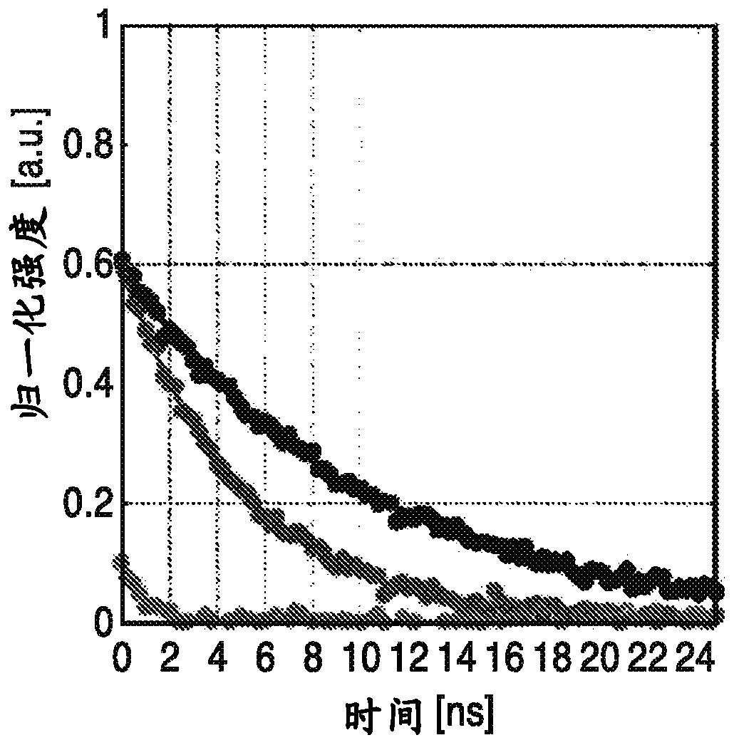
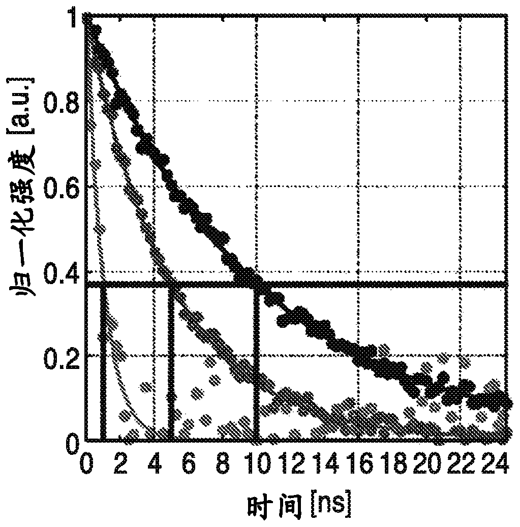
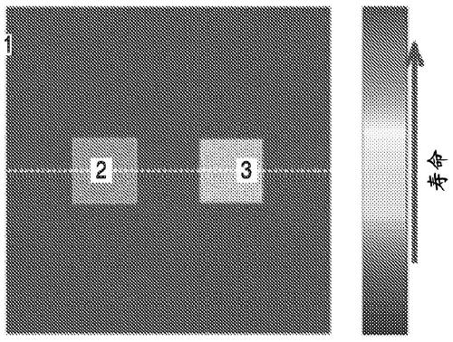
[0048] The systems and methods of the present specification take advantage of naturally occurring differences in fluorophore lifetimes between cell populations with different physiological processes for generating contrast, and apply unique algorithms to relax technical requirements.

[0049] For tissue autofluorescence, naturally occurring fluorophores are used to create contrast (eg, black light imaging). In one embodiment, the target is illuminated with short pulses of light and the emission intensity is measured as the emission decays from bright to dark. How long an area "glows" depends on what type of tissue is illuminated. For example, cancerous tissue is often associated with rapid decay, while non-cancerous tissue is associated with slow decay.

[0050] The systems and methods disclosed herein are configured for edge detection between populations of cells with different physiological processes, or edge detection between different tissues, such as but not limited to p...


PUM


Abstract

An imaging system and method are disclosed for intraoperative surgical margin assessment in between various tissues and cell groupings having differing physiologic processes. The system uses an arrayof LED's to pump a target anatomy with a short excitation pulse and measures the lifetime of fluorescence to generate contrast. A relative fluorescence lifetime map is generated corresponding to the measured lifetime to identify boundaries within varying cell groupings and tissues.

Description

[0001] Cross References to Related Applications [0002] This application claims priority to and benefit of U.S. Provisional Patent Application No. 62 / 580,383, filed November 1, 2017, the entire contents of which are hereby incorporated by reference. [0003] Statement Regarding Federally Funded Research and Development [0004] Notice of Non-Applicability to Copyrighted Material [0005] Portions of the material in this patent document may be protected by copyright under the copyright laws of the United States and other countries. The copyright owner has no objection to the facsimile reproduction by anyone of the patent document or the patent disclosure as it appears in publicly available documents or records of the USPTO, but otherwise reserves all copyrights. The copyright owner hereby does not waive any of its rights to keep this patent document confidential, including but not limited to rights under 37 CFR Section 1.14. [0006] background [0007] 1. Technical field ...

Claims


Application Information

Patent Timeline
Patent Type & Authority Applications(China)
IPC IPC(8): G01N33/483G01N21/64G01N21/27
CPCA61B5/0071A61B5/0077A61B5/444A61B5/4887G01N21/27G01N21/64G01N21/6408A61B90/39A61B2090/3904A61B2090/3941A61B5/0082A61B5/4866A61B2505/05A61B2560/0223A61B2562/0233A61B2562/04
Inventor 马耶·圣约翰乔治·萨迪克扎卡里·泰勒沃伦·格伦德费斯特
Owner RGT UNIV OF CALIFORNIA
Learn More