Method, device, terminal and storage medium for segmenting mammography image

An image segmentation and image technology, applied in image analysis, image enhancement, image data processing, etc., can solve the problems of rough binary image, unfavorable front-end display of breast mass and calcification research, and achieve the effect of improving the accuracy of breast segmentation

Active Publication Date: 2021-05-25
INFERVISION MEDICAL TECH CO LTD
View PDF3 Cites 0 Cited by
  • Summary
  • Abstract
  • Description
  • Claims
  • Application Information

AI Technical Summary

Problems solved by technology

The shortcomings of the existing methods include at least: the binary image obtained by segmentation network segmentation is relatively rough, which is not conducive to front-end display and subsequent research on breast masses and calcifications

Method used


Image

  • Method, device, terminal and storage medium for segmenting mammography image
  • Method, device, terminal and storage medium for segmenting mammography image
  • Method, device, terminal and storage medium for segmenting mammography image

Examples


Embodiment 1

[0040] figure 1 It is a schematic flow chart of a method for segmenting a mammography image provided in Embodiment 1 of the present invention. This embodiment is applicable to the case of performing breast region segmentation on a mammography image. The method can be performed by the segmentation device for mammography image provided by the embodiment of the present invention (may be referred to simply as the segmentation device), and the segmentation device can be configured in the terminal provided by the embodiment of the present invention, for example, it can be configured in conjunction with the mammography image In the computer equipment that communicates with the scanning equipment of the mammogram, or integrated in the scanning equipment of the mammogram image, no specific limitation is set here.

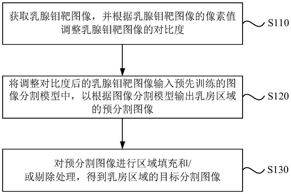
[0041] see figure 1 , a method for segmenting mammography images, specifically comprising the following steps:

[0042] S110. Acquire a mammography image, and adjust the c...

Embodiment 2

[0060] image 3 It is a schematic flow chart of a method for segmenting mammography images provided by Embodiment 2 of the present invention. On the basis of the above embodiments, this embodiment optimizes the training process when the image segmentation model is a convolutional neural network, which can improve Accuracy of Breast Region Segmentation. The methods for segmenting mammography images provided by the embodiments of the present invention and the above-mentioned embodiments belong to the same inventive concept, and the technical details not described in detail can be referred to the above-mentioned embodiments, and have the same technical effect.

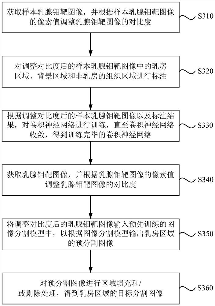
[0061] see image 3 , a method for segmenting mammography images, specifically comprising the following steps:

[0062] S310. Acquire a sample mammography image, and adjust the contrast of the mammography image according to the pixel values ​​of the sample mammography image.

[0063] In the embodiment of the present in...

Embodiment 3

[0085] Figure 5 It is a schematic structural diagram of a mammography image segmentation device provided in Embodiment 3 of the present invention. This embodiment is applicable to the case of performing breast region segmentation on a mammography image. The method for segmenting mammogram images provided by any embodiment of the present invention can be realized by using the device for segmenting mammogram images.

[0086] see Figure 5 , a segmentation device for mammography images, comprising:

[0087] The pre-processing module 510 is configured to acquire mammogram images, and adjust the contrast of the mammogram images according to the pixel values ​​of the mammogram images;

[0088] The pre-segmentation module 520 is used for inputting the contrast-adjusted mammary gland image into the pre-trained image segmentation model, so as to output the pre-segmentation image of the breast region according to the image segmentation model;

[0089] The post-processing module 530 ...


PUM


Abstract

The embodiment of the present invention discloses a segmentation method, device, terminal and storage medium of a mammogram image. The method includes: acquiring a mammogram image, and adjusting the contrast of the mammogram image according to the pixel values ​​of the mammogram image; Input the contrast-adjusted mammography image into the pre-trained image segmentation model to output the pre-segmented image of the breast region according to the image segmentation model; perform region filling and / or elimination processing on the pre-segmented image to obtain the target segmentation of the breast region image. By adjusting the contrast of the mammography image, the difference between the background area and the foreground area (breast area and non-breast tissue area) can be highlighted; the pre-segmentation of the breast area in the mammography image can be realized through the pre-trained image segmentation model; through Performing region filling and / or culling processing on pre-segmented images can improve the accuracy of breast segmentation, which is beneficial to front-end display and subsequent research on breast masses and calcifications.

Description

technical field [0001] Embodiments of the present invention relate to image segmentation technologies, and in particular to a segmentation method, device, terminal and storage medium for mammography images. Background technique [0002] At present, mammography examination, also known as molybdenum-palladium examination, uses low-dose X-rays to scan the breast, and obtains high-resolution mammography images through breast tissue absorbing X-rays to varying degrees. By performing breast segmentation on the mammography image, the information of the region of interest in the image can be determined, for example, the type of the region of interest is determined as calcification or mass. Compared with artificial breast examination, more complete and accurate breast information can be obtained through mammography, and the examination method is simple, reliable, non-invasive, reproducible, not limited by age and body shape, so it has been used as a routine breast examination means....

Claims


Application Information

Patent Timeline
Patent Type & Authority Patents(China)
IPC IPC(8): G06T7/00G06T7/11G06T7/194G06T7/62G06T5/00G06N3/04G06N3/08
CPCG06T7/0012G06T7/11G06T7/194G06T7/62G06T5/009G06N3/08G06T2207/10081G06T2207/30068G06N3/045
Inventor 黄文豪赵朝炜王瑜周越孙岩峰邹彤李新阳王少康陈宽
Owner INFERVISION MEDICAL TECH CO LTD
Why Eureka
Try Eureka