
Portable device for removing ureteral stent through urethra

A portable device, ureteral technology, applied in urethoscope, application, endoscope and other directions, can solve the problems of cross infection, increase the economic burden of patients, and high cost, achieve no pollution, facilitate continuous surgical operation, and low cost and price Effect

Pending Publication Date: 2020-10-13
湘雅常德医院
View PDF0 Cites 0 Cited by
  • Summary
  • Abstract
  • Description
  • Claims
  • Application Information

AI Technical Summary

Problems solved by technology

The working channel of the foreign body forceps is on the cystoscope (integrated with the cystoscope). The foreign body forceps enters the bladder through this working channel to grab the ureteral stent tube. This instrument (cystoscope and foreign body forceps) is repetitive The instruments used need to be completely and effectively sterilized and aseptically packaged after each use, and this operation needs to be completed in the operating room or cystoscopy room, which is costly and has the possibility of cross-infection. In addition, because After each use, there is a requirement for disinfection and sterilization, so surgery scheduling cannot be performed continuously, that is, a cystoscope can only be used once in a short period of time, especially in overcrowded large hospitals, patients may have to wait for the next day before removing the cystoscope, increasing the Patient's Financial Burden

Method used


Image

  • Portable device for removing ureteral stent through urethra
  • Portable device for removing ureteral stent through urethra
  • Portable device for removing ureteral stent through urethra

Examples


Embodiment Construction

[0016] The present invention will be described in further detail below in conjunction with the accompanying drawings and embodiments.

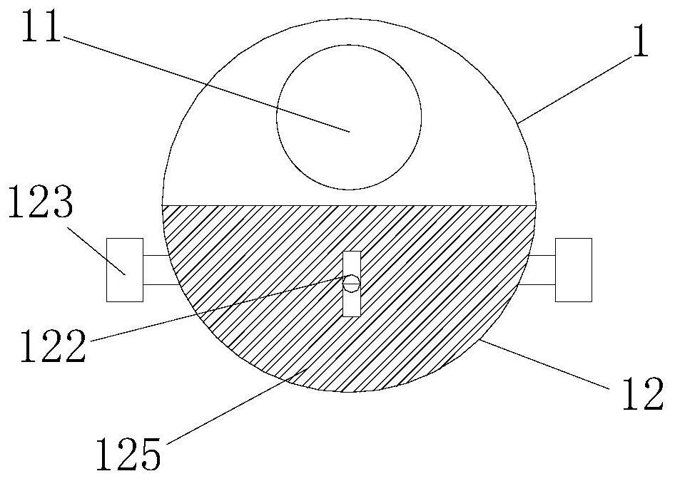
[0017] Refer to attached Figure 1-3 , the present embodiment includes a visible fiberscope sleeve 1, a visible fiberscope 2 sleeved in the visible fiberscope sleeve 1, the fiberscope sleeve 1 is provided with two independent fiberscope channels 11 and 12 working channels The visible fiberscope 2 is arranged in the fiberscope channel 11, and the front end 111 of the fiberscope channel 11 is a blind end and is transparently sealed, which does not prevent the optical fiber from producing a surgical field of view, and does not have any contact with human tissue; The front end 121 of the working channel 12 has the same structure as the front end of the existing ureteroscope, and the working channel 12 is also provided with a spring clip 122, and the spring clip 122 can enter the human bladder 3 through the working channel 12, and pull out the uret...


PUM


Abstract

Disclosed is a portable device for removing a ureteral stent through urethra. The portable device comprises a visible fiber optic endoscope sleeve and a visible fiber optic endoscope sleeved with thevisible fiber optic endoscope sleeve, wherein the fiber optic endoscope sleeve is provided with two independent channels including a fiber optic endoscope channel and a working channel; the visible fiber optic endoscope is arranged in the fiber optic endoscope channel; the front end of the optical fiber endoscope channel is a blind end and is transparent, sealed and capable of guiding light; the front end of the working channel and the front end of a ureteroscope are of the same structure; and a spring clamp is further arranged in the working channel, and the spring clamp can enter the bladderof a human body through the working channel. By means of the portable device, the visible optical fiber endoscope can be repeatedly used after an operation without sterilization treatment, on the premise that the optical fiber does not hinder the operation view field, the visible optical fiber endoscope does not make contact with human tissue at all, and therefore the portable device can meet thedisposable use requirement, is low in cost and price, does not generate pollution and facilitates continuous operation.

Description

technical field [0001] The invention relates to a medical instrument device, in particular to a portable device for pulling out a ureteral stent through the urethra. Background technique [0002] Clinically, after surgery for ureteral stricture and upper urinary tract stones, ureteral stents are routinely indwelled to play the role of expansion, support and drainage after surgery. Usually about 1-3 months, the foreign body forceps are pulled out under the cystoscope. The working channel of the foreign body forceps is on the cystoscope (integrated with the cystoscope). The foreign body forceps enters the bladder through this working channel to grab the ureteral stent tube. This instrument (cystoscope and foreign body forceps) is repetitive The instruments used need to be completely and effectively sterilized and aseptically packaged after each use, and this operation needs to be completed in the operating room or cystoscopy room, which is costly and has the possibility of cr...

Claims


Application Information

Patent Timeline
Patent Type & Authority Applications(China)
IPC IPC(8): A61B1/307A61B1/018A61B1/04A61B1/06A61B1/07A61B1/00A61B17/00A61B17/50
CPCA61B1/00165A61B1/018A61B1/04A61B1/0684A61B1/07A61B1/307A61B17/00234A61B17/50A61B2017/00353
Inventor 王维国郑若兰夏菲蒋欣杰李国军陈诚陈旭东顿金庚
Owner 湘雅常德医院
Learn More