Multi-ring collimator imaging structure and imaging device of full-ring SPECT

A collimator and imaging technology, applied in the field of collimators, can solve the problems of poor patient inspection experience, safety risks, and low inspection efficiency.

Pending Publication Date: 2020-10-23
ATOMICAL MEDICAL EQUIP FO SHAN LTD
View PDF0 Cites 2 Cited by
  • Summary
  • Abstract
  • Description
  • Claims
  • Application Information

AI Technical Summary

Problems solved by technology

[0002] The existing SPECT collimator probes are mostly dual-probe collimators with arms stretched out. The inspection function of the collimator is single. For different organ inspections, the corresponding collimators need to be replaced manually or automatically. The probe cannot fully cover the main organs of the body. During the inspection process, the angle, position and other parameters of the probe need to be adjusted for different organ inspections. Therefore, if multiple organs are used for SPECT, the whole body inspection will take a long time and the inspection efficiency will be low. disadvantage
[0003] At the same t

Method used


Image

  • Multi-ring collimator imaging structure and imaging device of full-ring SPECT
  • Multi-ring collimator imaging structure and imaging device of full-ring SPECT
  • Multi-ring collimator imaging structure and imaging device of full-ring SPECT

Examples


Embodiment Construction

[0040] The technical solutions of the present invention will be further described below through specific implementations with reference to the drawings.

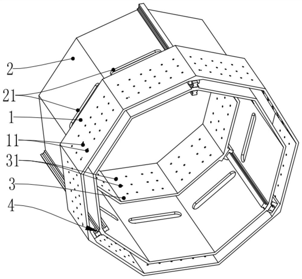
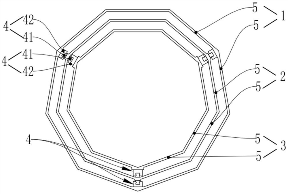
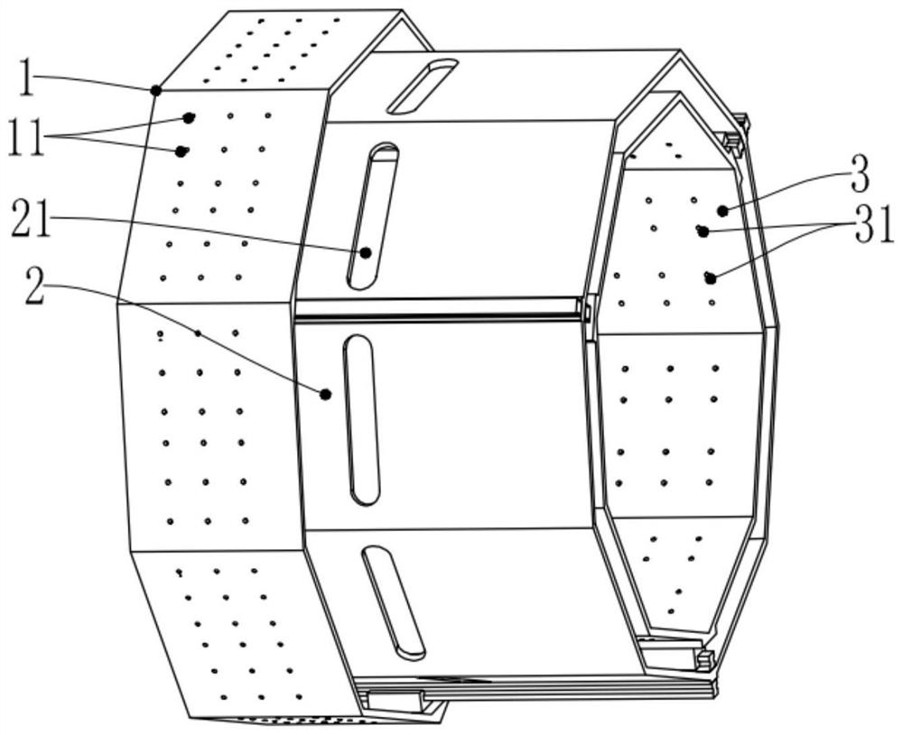
[0041] A multi-ring collimator imaging structure for full-ring SPECT, including: a pinhole outer ring 1, a fully shielded middle ring 2 and a pinhole inner ring 3;

[0042] The surface of the pinhole outer ring 1 is provided with a pin outer hole 11; the surface of the pinhole inner ring 3 is provided with a pin inner hole 31; the surface of the fully shielded intermediate ring 2 is provided with a ray penetrating port 21;

[0043] The pinhole outer ring 1 can be relatively movably installed outside the ring of the fully shielded intermediate ring 2, so that when the pinhole outer ring 1 passes through the ray penetration opening 21, the pin outer hole 11 passes through and Aligned to the ray penetration opening 21;

[0044] The pinhole inner ring 3 can be relatively movably installed in the ring of the fully shielded intermediate r...


PUM


Abstract

The invention discloses a multi-ring collimator imaging structure and imaging device of full-ring SPECT. The surface of an outer ring of a pinhole of the multi-ring collimator imaging structure is provided with a needle outer hole; a needle inner hole is formed in the surface of an inner ring of the pinhole; a ray penetrating opening is formed in the surface of a full-shielding middle ring; the pinhole outer ring can be relatively movably mounted outside the full-shielding middle ring, so that when the pinhole outer ring passes through the ray penetration opening, the pinhole outer ring passesthrough and is aligned with the ray penetration opening; and the pinhole inner ring can be relatively movably arranged in the ring of the full-shielding middle ring, so that when the pinhole inner ring passes through the ray penetration opening, the needle inner hole passes through and is aligned with the ray penetration opening. According to the multi-ring collimator imaging device, the pinholeouter ring, the full-shielding middle ring and the pinhole inner ring form an imaging unit. According to the scheme, the collimator does not need to be replaced, redundant rays are shielded, only therays emitted by the inspected organ and the pinhole are reserved, and the function of automatically adapting to the inspected organ in a high-precision mode is achieved, so that the inspection efficiency is greatly improved, the inspection time is shortened, the imaging quality is improved, and the inspected organ is accurately positioned.

Description

Technical field [0001] The present invention relates to the technical field of collimators, in particular to a multi-ring collimator imaging structure and imaging device for full-ring SPECT. Background technique [0002] Existing SPECT collimator probes are mostly dual-probe collimators with arms outstretched. The collimator has a single inspection function. For different organ inspections, the corresponding collimator needs to be replaced manually or automatically. In addition, the double The probe cannot fully cover the main organs of the body. During the inspection process, it is necessary to adjust the probe angle, position and other parameters for different organs. Therefore, if there are multiple organs in SPECT, the whole body inspection takes longer and the inspection efficiency is lower. Disadvantage. [0003] At the same time, the existing dual SPECT probes cannot cover the entire human body, resulting in relatively blurred imaging quality. Moreover, the double probe it...

Claims


Application Information

Patent Timeline
IPC IPC(8): A61B6/03G21K1/02
CPCA61B6/037A61B6/4429A61B6/542G21K1/02
Inventor 谢振华王维民邓晓陈思
Owner ATOMICAL MEDICAL EQUIP FO SHAN LTD
Why Eureka
Try Eureka