Image reconstruction method and application thereof

An image reconstruction and image technology, which is applied in the field of image scanning, can solve the problems of radiation lesions in the scanned parts, large X-ray radiation doses of patients, etc., to achieve reduced radiation doses, high imaging quality, and avoid reconstructed image artifacts and wrong information problem effect

Pending Publication Date: 2020-10-30
国创育成医疗器械发展(深圳)有限公司
View PDF0 Cites 0 Cited by
  • Summary
  • Abstract
  • Description
  • Claims
  • Application Information

AI Technical Summary

Problems solved by technology

[0006] Based on existing image reconstruction, in order to reconstruct a dynamic image sequence that meets clinical diagnostic standards, it is necessary to repeat multiple CT scans of the same dose on the patient, which will bring a large X-ray radiation dose to the patient and have potential In view of the problem of the risk of radiation lesions in the scanned part, this application provides an image reconstruction method and its application

Method used


Image

  • Image reconstruction method and application thereof
  • Image reconstruction method and application thereof
  • Image reconstruction method and application thereof

Examples


Embodiment

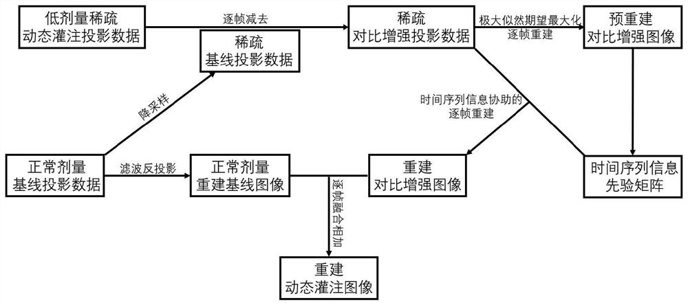
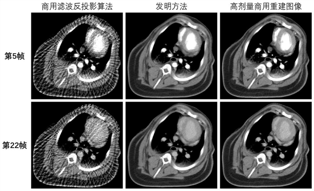
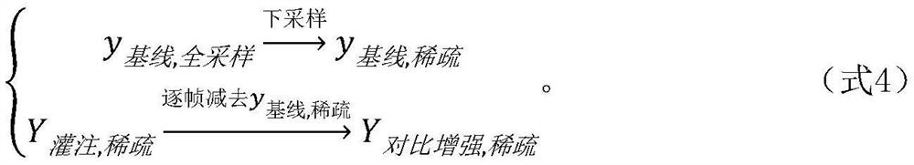
[0062] Based on the separation and reconstruction method of dynamic perfusion-enhanced CT images and the sparse angle image acquisition scheme of dynamic image sequences, the contrast-enhanced images of cardiac tissue caused by imaging agents are reconstructed with the help of the time evolution information of imaging agents naturally contained in dynamic image sequences, The reconstructed enhanced image was superimposed on the baseline CT image obtained by the normal dose acquisition scheme to obtain the final dynamic myocardial perfusion scanning CT image sequence.

[0063] Image data acquisition: baseline image acquisition for myocardial dynamic perfusion CT imaging, that is, the CT scan performed before the imaging agent reaches the human heart tissue, and dynamic perfusion image sequence acquisition, that is, the imaging agent performed after the imaging agent reaches the human heart tissue Consecutive CT scans, with different doses of CT image acquisition schemes. The ba...


PUM


Abstract

The invention belongs to the technical field of image scanning, and particularly relates to an image reconstruction method and application thereof. Existing image reconstruction needs to perform repeated same-dose CT scanning on a patient for many times to reconstruct a dynamic image sequence meeting clinical diagnosis standards, so a large X-ray radiation dose is brought to the patient, and a potential risk of causing radiation lesion of a scanned part exists. The invention discloses a separation and reconstruction method based on a dynamic perfusion enhanced image and a sparse angle image acquisition scheme based on a dynamic image sequence. The method comprises the steps: reconstructing a heart tissue contrast enhanced image caused by a developer by means of developer time evolution information naturally contained in the dynamic image sequence, and superposing the reconstructed enhanced image to a baseline image obtained by a normal dose acquisition scheme to obtain a final dynamicperfusion scanning image sequence. And the radiation dose of dynamic perfusion imaging on a patient is remarkably reduced.

Description

technical field [0001] The present application belongs to the technical field of image scanning, and in particular relates to an image reconstruction method and its application. Background technique [0002] Image reconstruction is a technology that obtains the shape information of a three-dimensional object through digital processing of the data measured outside the object. Image reconstruction technology was initially applied in radiation medical equipment to display images of various parts of the human body, that is, computed tomography technology, or CT technology for short, and has gradually been applied in many fields. There are mainly projection reconstruction, shading recovery shape, stereo vision reconstruction and laser ranging reconstruction. [0003] Dynamic myocardial perfusion CT imaging (DMP-CT) is an effective examination method for clinical diagnosis of coronary artery disease. Existing clinical DMP-CT imaging requires repeated CT scanning of the patient a...

Claims


Application Information

Patent Timeline
Patent Type & Authority Applications(China)
IPC IPC(8): G06T11/00
CPCG06T11/003G06T2211/416G06T2211/421
Inventor 李彦明郑海荣江洪伟万丽雯
Owner 国创育成医疗器械发展(深圳)有限公司
Why Eureka
Try Eureka