
Automatic ventricular region segmentation method and device of cardiac ultrasound image and storage medium

An automatic segmentation and ultrasound image technology, applied in the field of image processing, can solve the problems of model training results failing to converge, obvious echocardiographic noise, fixed position of the region to be segmented, etc., to achieve easy deployment, improve training speed and accuracy, improve The effect of speed and accuracy

Pending Publication Date: 2020-11-10
东软教育科技集团有限公司
View PDF5 Cites 2 Cited by
  • Summary
  • Abstract
  • Description
  • Claims
  • Application Information

AI Technical Summary

Problems solved by technology

[0004] 1) Echocardiography has obvious noise, and the position of the region to be segmented is relatively fixed.
Existing segmentation techniques using neural networks do not express enough information from multiple scales, and there is still much room for improvement
[0005] 2) At present, the optimization objective function of the image segmentation model training still uses the traditional MSE (mean square error function), which will make the model training results unable to converge

Method used


Image

  • Automatic ventricular region segmentation method and device of cardiac ultrasound image and storage medium
  • Automatic ventricular region segmentation method and device of cardiac ultrasound image and storage medium
  • Automatic ventricular region segmentation method and device of cardiac ultrasound image and storage medium

Examples


Embodiment Construction

[0054] In order to enable those skilled in the art to better understand the solutions of the present invention, the following will clearly and completely describe the technical solutions in the embodiments of the present invention in conjunction with the drawings in the embodiments of the present invention. Obviously, the described embodiments are only It is an embodiment of a part of the present invention, but not all embodiments. Based on the embodiments of the present invention, all other embodiments obtained by persons of ordinary skill in the art without making creative efforts shall fall within the protection scope of the present invention.

[0055] It should be noted that the terms "first" and "second" in the description and claims of the present invention and the above drawings are used to distinguish similar objects, but not necessarily used to describe a specific sequence or sequence. It is to be understood that the data so used are interchangeable under appropriate ...


PUM


Abstract

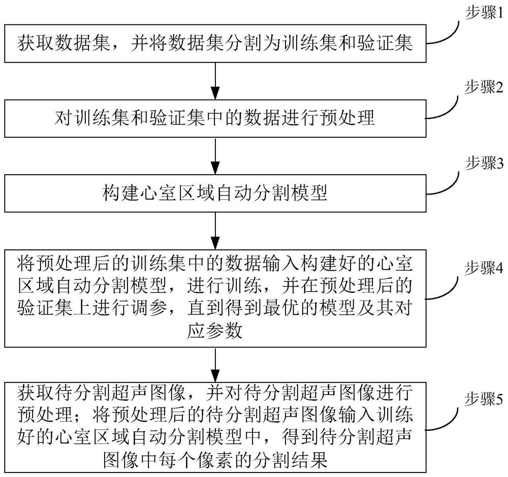
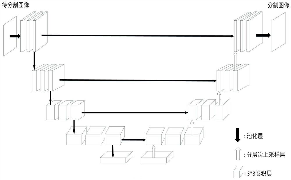
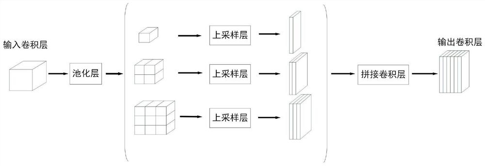
The invention provides an automatic ventricular region segmentation method and device for a cardiac ultrasound image and a storage medium, and relates to the technical field of image processing. According to the method, a ventricular region automatic segmentation model is utilized to obtain a segmentation result of each pixel in a to-be-segmented ultrasonic image. According to the ventricular region automatic segmentation model, compared with a traditional UNet method, the network structure of the model is improved, an up-sampling deconvolution layer of a decoder of the model is a multi-dimensional splicing type deconvolution up-sampling layer, the decoder can better obtain image information from multiple dimensions, and then the segmentation accuracy is improved. According to the automatic ventricular region segmentation model, the model training speed and accuracy can be effectively improved, and then the speed and accuracy of automatically segmenting the left ventricular region in the echocardiogram are improved.

Description

technical field [0001] The present invention relates to the technical field of image processing, in particular to a method, device and storage medium for automatic segmentation of ventricle regions of cardiac ultrasound images. Background technique [0002] Cardiac ejection fraction is an important indicator for judging the type of heart failure and one of the important methods for measuring heart function. Cardiac ejection fraction refers specifically to the percentage of blood that the heart ejects during systole after filling with blood during diastole. The first step to obtain this index relies on the segmentation of the ventricles of the heart image. [0003] In recent years, many different methods have been proposed and used for the automatic segmentation of cardiac images, which are mainly divided into two categories from the general direction: one is the expansion and improvement of traditional segmentation algorithms; the other is the combination of deep learning a...

Claims


Application Information

Patent Timeline
Patent Type & Authority Applications(China)
IPC IPC(8): G06T7/11G06N3/04G06T3/40G06K9/62G06K9/32
CPCG06T7/11G06T3/4084G06T2207/20081G06T2207/20084G06T2207/30048G06V10/25G06N3/045G06F18/214
Inventor 包一平张宾于丹李雪
Owner 东软教育科技集团有限公司
Learn More