Brain white matter region segmentation method and device for craniocerebral ultrasonic image and electronic equipment

A technology for ultrasonic image and region segmentation, applied in the field of image processing, which can solve the problems of negative impact on segmentation results and low proportion of brain white matter regions

Pending Publication Date: 2020-11-17
SHENZHEN INST OF ADVANCED TECH CHINESE ACAD OF SCI
View PDF0 Cites 1 Cited by
  • Summary
  • Abstract
  • Description
  • Claims
  • Application Information

AI Technical Summary

Problems solved by technology

[0004] The purpose of the present invention is to provide a brain white matter region segmentation method for craniocerebral ultrasound images, aiming to solve the techni...

Method used


Image

  • Brain white matter region segmentation method and device for craniocerebral ultrasonic image and electronic equipment
  • Brain white matter region segmentation method and device for craniocerebral ultrasonic image and electronic equipment
  • Brain white matter region segmentation method and device for craniocerebral ultrasonic image and electronic equipment

Examples


Embodiment 1

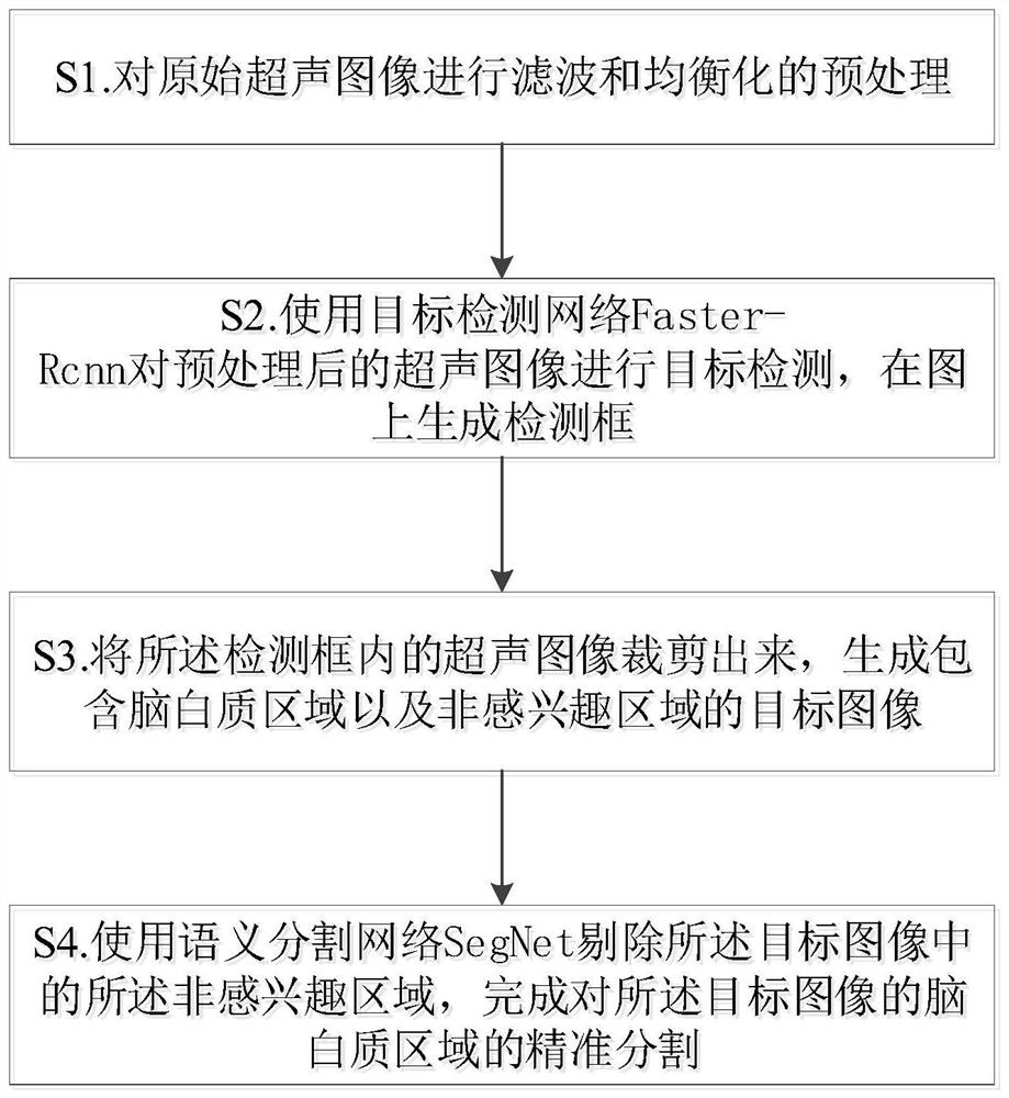
[0027] figure 1 , image 3 It shows a method for segmenting brain white matter regions of a craniocerebral ultrasound image provided in the first embodiment of the present invention, and the method includes the following steps:

[0028] S1. Perform filtering and equalization preprocessing on the original ultrasound image;

[0029] S2. Use the target detection network Faster-Rcnn to perform target detection on the preprocessed ultrasound image, and generate a detection frame on the image;

[0030] S3. Cut out the ultrasound image in the detection frame to generate a target image including the white matter region and the region of non-interest;

[0031] S4. Use the semantic segmentation network SegNet to eliminate the non-interest region in the target image, and complete the accurate segmentation of the white matter region of the target image.

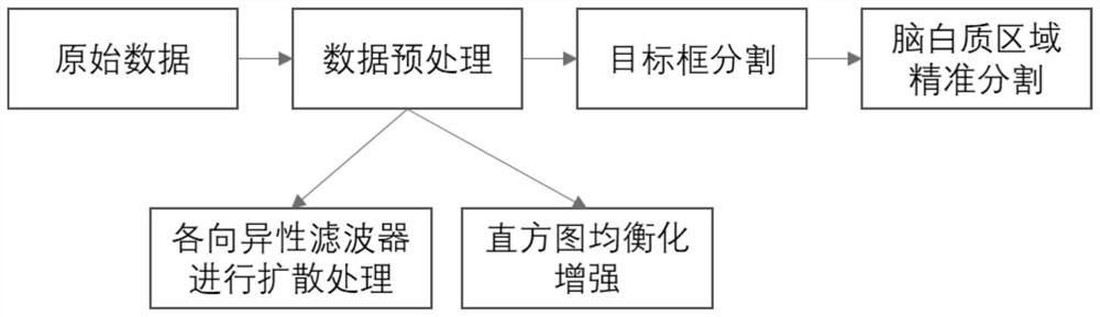
[0032] Further, the step S1 includes the following steps:

[0033] S11. Use an anisotropic filter to perform diffusion processing on the original ...

Embodiment 2

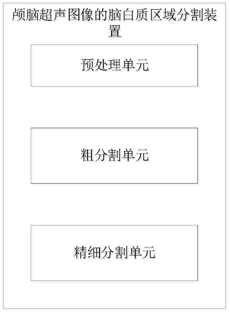
[0054] figure 2 It shows a brain white matter region segmentation device for craniocerebral ultrasound images provided in the second embodiment of the present invention, including:

[0055] The preprocessing unit is used for filtering and equalizing the preprocessing of the original ultrasound image;

[0056] The coarse segmentation unit uses the target detection network Faster-Rcnn to perform target detection on the pre-processed ultrasound image, and generates a detection frame on the image; then, the ultrasound image in the detection frame is cropped out to generate regions containing white matter and non-sensing The target image of the region of interest;

[0057] The fine segmentation unit uses the semantic segmentation network SegNet to eliminate the non-interest region in the target image, and complete the precise segmentation of the white matter region of the target image.

[0058] Further, the preprocessing unit includes:

[0059] A filtering module, which uses an anisotropic...

Embodiment 3

[0072] An electronic device provided in the third embodiment of the present invention includes:

[0073] At least one processor; and

[0074] A memory communicatively connected to the at least one processor; the memory stores instructions executable by the one processor, and the instructions are executed by the at least one processor to enable the at least one processor to execute The brain white matter region segmentation method for craniocerebral ultrasound images as described in any one of the above.

[0075] The various drawbacks of manual segmentation are solved, and the automatic segmentation method is adopted to reduce the burden on doctors.


PUM


Abstract

The invention is applicable to the technical field of image processing, and provides a brain white matter region segmentation method and device for a craniocerebral ultrasonic image and electronic equipment. The method comprises the following steps: carrying out diffusion and enhancement on an original input ultrasonic image, and then carrying out rough segmentation on the processed ultrasonic image to obtain a target image containing a brain white matter area and other non-interested areas; and carrying out secondary segmentation on a narrowed area, and removing the non-interested areas in the frame to obtain an accurately segmented brain white matter area. The invention can effectively avoid the problems that there is no clear boundary between an interested area and the surrounding environment in an ultrasonic image, the proportion of a brain white matter area is too low, and veins and other highlight areas have very strong negative effects on a segmentation result. Meanwhile, the white matter area segmentation device of the craniocerebral ultrasonic image and the electronic equipment provided by the invention can also achieve the same technical effect.

Description

Technical field [0001] The invention belongs to the technical field of image processing, and in particular relates to a brain white matter region segmentation method, device and electronic equipment of craniocerebral ultrasound images. Background technique [0002] Traditional white matter ultrasound image analysis methods use manual segmentation. If the damaged parts of the brain white matter are the anterior horn of the lateral ventricle, the white matter near the posterior horn, the lateral and dorsal white matter of the lateral ventricle, or the subcortical white matter, the accuracy of manual segmentation is likely to be affected by the operator's experience level and cause artificial errors. In addition, traditional methods perform feature extraction and classification after manual segmentation, which consumes a lot of manpower and material resources. [0003] The difficulty in segmentation of the white matter region of ultrasound images is that the image contrast is low, es...

Claims


Application Information

Patent Timeline
IPC IPC(8): G06T7/11G06T5/40G06K9/32G06K9/62G06N3/04
CPCG06T7/11G06T5/40G06T2207/10132G06T2207/20132G06T2207/20081G06T2207/20084G06T2207/30016G06V10/25G06V2201/03G06V2201/07G06N3/045G06F18/241
Inventor 张湘楠陈智毅廖剑艺梁晓雯
Owner SHENZHEN INST OF ADVANCED TECH CHINESE ACAD OF SCI
Why Eureka
Try Eureka