Three-dimensional visualization preoperative planning method, system and terminal for abdominal wall defect reconstruction

An abdominal wall, three-dimensional technology, applied in the medical field, can solve the problems of not being able to display the spatial position relationship of the lesions in three-dimensional, increasing the error of cutting operation, wasting time and manpower, etc., so as to reduce the operation time, improve the success rate of the operation, and reduce the postoperative complications. The effect of disease incidence

Active Publication Date: 2021-09-21
SHANGHAI NINTH PEOPLES HOSPITAL SHANGHAI JIAO TONG UNIV SCHOOL OF MEDICINE
View PDF7 Cites 0 Cited by
  • Summary
  • Abstract
  • Description
  • Claims
  • Application Information

AI Technical Summary

Problems solved by technology

[0003] In view of the above-mentioned shortcomings of the prior art, the purpose of the present invention is to provide a three-dimensional visualized preoperative planning method, system and terminal for abdominal wall defect reconstruction, which is used to solve the problem of inability to three-dimensionally display the lesion and the surrounding adjacent blood vessels in the prior art. The spatial positional relationship of tissues, organs, and muscles has caused great difficulties for doctors to formulate and simulate surgical plans, and manual segmentation of large-scale clinical data will not only waste a lot of time and manpower, but also increase the error of cutting operations, and further Greatly reduce the efficiency and accuracy of cutting operations

Method used


Image

  • Three-dimensional visualization preoperative planning method, system and terminal for abdominal wall defect reconstruction
  • Three-dimensional visualization preoperative planning method, system and terminal for abdominal wall defect reconstruction
  • Three-dimensional visualization preoperative planning method, system and terminal for abdominal wall defect reconstruction

Examples


Embodiment 1

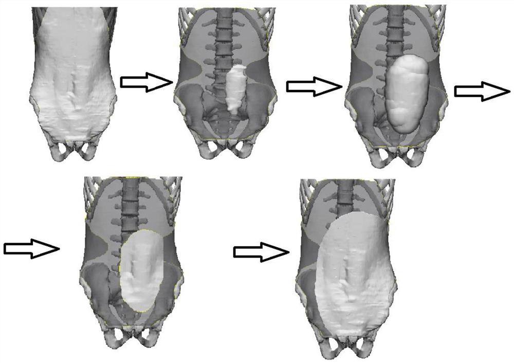
[0064] Example 1: A three-dimensional visualization preoperative planning method for abdominal wall defect reconstruction. Figure 3 is a schematic diagram of the state of the abdominal wall during the three-dimensional patch acquisition process.

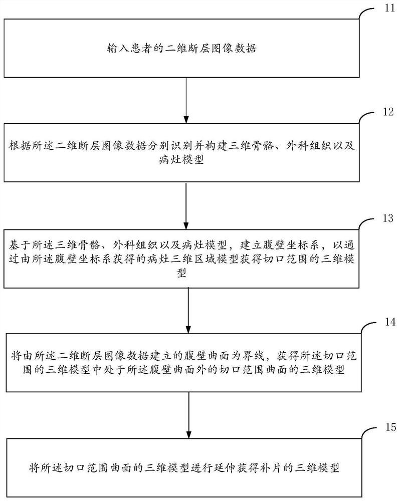
[0065] The methods include:

[0066]Input the two-dimensional tomographic image data of the patient; identify the three-dimensional bones, surgical tissues and lesions in the image data according to the input two-dimensional tomographic image, and construct the three-dimensional bones, surgical tissues and lesions models, and establish an abdominal wall coordinate system, A three-dimensional model of the incision range is obtained from the three-dimensional region model of the lesion obtained from the abdominal wall coordinate system. Perform curved surface reconstruction according to the two-dimensional tomographic image data to obtain a curved surface of the abdominal wall; with the curved surface of the abdomen as a limit, calcul...

specific Embodiment

[0069] Such as Figure 4 A schematic structural diagram of a system showing a three-dimensional visualized preoperative planning method for abdominal wall defect reconstruction in an embodiment of the present invention.

[0070] The system includes:

[0071] A two-dimensional tomographic image input module 41, configured to input two-dimensional tomographic image data of the patient;

[0072] A three-dimensional model construction module 42, connected to the two-dimensional tomographic image input module 41, for identifying and constructing three-dimensional bone, surgical tissue and lesion models respectively according to the two-dimensional tomographic image data;

[0073] The incision range module 43 is connected to the three-dimensional model building module 42, and is used to establish an abdominal wall coordinate system based on the three-dimensional bone, surgical tissue and lesion models, so as to obtain the three-dimensional area of ​​the incision range through the t...


PUM


Abstract

The three-dimensional visualization preoperative planning method, system and terminal for abdominal wall defect reconstruction of the present invention solve the problem that the prior art cannot three-dimensionally display the spatial position relationship between the lesion and the surrounding adjacent blood vessels, tissues, organs, and muscles, and is helpful for doctors to formulate and simulate operations. The solution caused great difficulties, and manual segmentation wasted a lot of time and manpower, which greatly reduced the efficiency and accuracy of cutting operations. The present invention can not only accurately display the location, size, internal structure and relationship with surrounding tissues in the three-dimensional reconstruction image of abdominal wall lesions constructed based on two-dimensional data, but also simulate the expansion of abdominal wall lesions on the premise of avoiding damage to important anatomical structures Abdominal wall defect after resection and the resection of invading tissues and organs, planning the surgical process of patch placement and abdominal wall reconstruction can not only reduce the operation time, but also reduce the incidence of postoperative complications, which has a significant and positive effect on improving the success rate of surgery. effect.

Description

technical field [0001] The present invention relates to the medical field, in particular to a three-dimensional visualized preoperative planning method, system and terminal for abdominal wall defect reconstruction. Background technique [0002] CT and MRI scans are routine examinations for the diagnosis of abdominal-related diseases, such as pathological damage or changes in abdominal organs. However, qualitative evaluation of only CT or MRI 2D images is highly subjective. Therefore, quantitative analysis is very important for precise diagnosis and treatment, and some measurement indicators can quantitatively and systematically evaluate the severity of the disease, so that surgeons can take more appropriate treatment measures. The segmentation of two-dimensional images provides some characteristics of the target area, such as size, width, position and other properties, and quantitative analysis can be carried out on this basis. On the other hand, in some surgical operation...

Claims


Application Information

Patent Timeline
Patent Type & Authority Patents(China)
IPC IPC(8): A61B34/10
CPCA61B34/10A61B2034/105A61B2034/107
Inventor 顾岩杨建军宋致成汪轶平
Owner SHANGHAI NINTH PEOPLES HOSPITAL SHANGHAI JIAO TONG UNIV SCHOOL OF MEDICINE
Why Eureka
Try Eureka