
Spine detection method and device, electronic equipment and storage medium

A detection method and spine technology, applied in the field of medical image processing, can solve the problems of error in diagnosis results, low efficiency, and time and energy consumption of clinicians, and achieve the effect of improving work efficiency, reducing work burden, and saving manpower and time consumption.

Pending Publication Date: 2021-01-15
沈阳先进医疗设备技术孵化中心有限公司
View PDF0 Cites 13 Cited by
  • Summary
  • Abstract
  • Description
  • Claims
  • Application Information

AI Technical Summary

Problems solved by technology

In clinical practice, clinicians with rich theoretical knowledge and practical experience are usually required to manually measure scoliosis and spinal stenosis from patients' CT images, which consumes a lot of time and energy for clinicians and is inefficient
And affected by the knowledge accumulation, practical experience and subjective factors of different doctors, the diagnostic results obtained by different doctors will also have large errors

Method used


Image

  • Spine detection method and device, electronic equipment and storage medium
  • Spine detection method and device, electronic equipment and storage medium
  • Spine detection method and device, electronic equipment and storage medium

Examples


Embodiment Construction

[0039] Reference will now be made in detail to the exemplary embodiments, examples of which are illustrated in the accompanying drawings. When the following description refers to the accompanying drawings, the same numerals in different drawings refer to the same or similar elements unless otherwise indicated. The implementations described in the following exemplary examples do not represent all implementations consistent with the present invention. Rather, they are merely examples of apparatuses and methods consistent with aspects of the embodiments of the invention as recited in the appended claims.

[0040] Terms used in the embodiments of the present invention are only for the purpose of describing specific embodiments of the present invention, and are not intended to limit the embodiments of the present invention. As used in the embodiments of the present invention and the appended claims, the singular forms "a", "said" and "the" are also intended to include the plural f...


PUM


Abstract

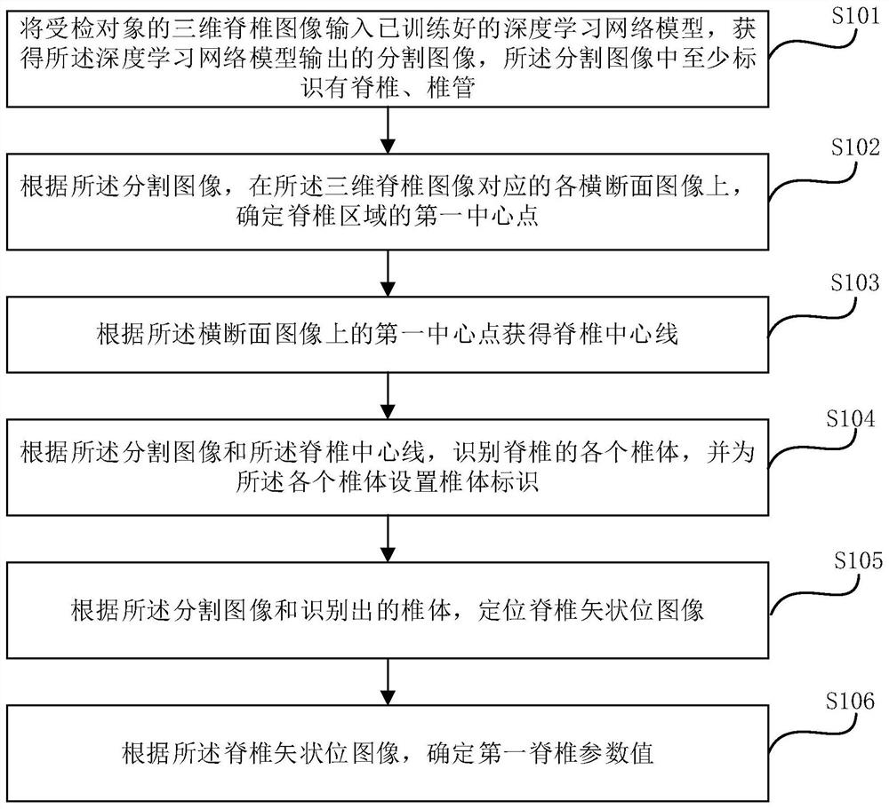
The embodiment of the invention provides a spine detection method and device, electronic equipment and a storage medium. According to the embodiment of the invention, the method comprises the steps: inputting a three-dimensional spine image into a deep learning network model, obtaining a segmented image outputted by the deep learning network model, and determining a first central point of a spineregion on each cross section image corresponding to the three-dimensional spine image according to the segmented image, obtaining a spine center line according to a first center point on the cross section image, identifying each spine body of the spine according to the segmentation image and the spine center line, setting a spine body identifier for each spine body, positioning a spine sagittal image according to the segmentation image and the identified spine body, and determining a first spinal parameter value according to the spine sagittal image. According to the method and device, the spine parameters can be measured in a full-automatic mode, and the working efficiency is improved.

Description

technical field [0001] The invention relates to the technical field of medical image processing, in particular to a vertebra detection method, device, electronic equipment and storage medium. Background technique [0002] With the continuous advancement of medical imaging technology, CT (Computed Tomography, computerized tomography technology) is widely used in the diagnosis of spinal stenosis. In clinical practice, clinical experts with rich theoretical knowledge and practical experience are usually required. Doctors manually measure scoliosis and spinal stenosis from the patient's CT images, which consumes a lot of time and energy for clinicians, and is inefficient. And affected by the accumulation of knowledge, practical experience and subjective factors of different doctors, the results measured by different doctors Diagnosis results will also have a large error. Contents of the invention [0003] In order to overcome the problems existing in the related technologies,...

Claims


Application Information

Patent Timeline
IPC IPC(8): G06T7/00G06T7/11G06T7/73G06N3/04G06N3/08
CPCG06T7/0012G06T7/11G06T7/73G06N3/08G06T2207/10081G06T2207/30012G06T2207/20081G06T2207/20084G06T2207/30204G06N3/045
Inventor 李丙生何薇
Owner 沈阳先进医疗设备技术孵化中心有限公司
Learn More