Liver CT image multi-lesion classification method based on sample generation and transfer learning

A CT image and transfer learning technology, applied in the field of image processing, can solve the problems of detection and classification of multiple types of lesions, and achieve the effect of improving lesion classification performance, accurate and fast lesion classification performance, and speeding up training.

Active Publication Date: 2021-01-19
XIDIAN UNIV
View PDF24 Cites 21 Cited by
  • Summary
  • Abstract
  • Description
  • Claims
  • Application Information

AI Technical Summary

Problems solved by technology

[0005] Among the current research methods for lesion detection and classification, the traditional image processing method through artificial design and lesion-related features, and the deep learning method through convolutional neural network learni

Method used


Image

  • Liver CT image multi-lesion classification method based on sample generation and transfer learning
  • Liver CT image multi-lesion classification method based on sample generation and transfer learning
  • Liver CT image multi-lesion classification method based on sample generation and transfer learning

Examples


Embodiment Construction

[0040] The present invention will be described in further detail below in conjunction with the accompanying drawings.

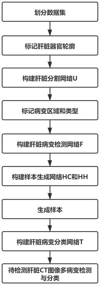
[0041] refer to figure 1 , the implementation steps of the present invention are as follows:

[0042] Step 1. Divide the dataset, mark the outline of the liver organ and store it.

[0043] 1.1) Extract 420 2D liver CT images from the liver CT images, and randomly select 80% of the images as a training set, and the remaining 20% ​​as a test set;

[0044] 1.2) Mark the contours of liver organs on all 2D liver CT images and store them as mask images.

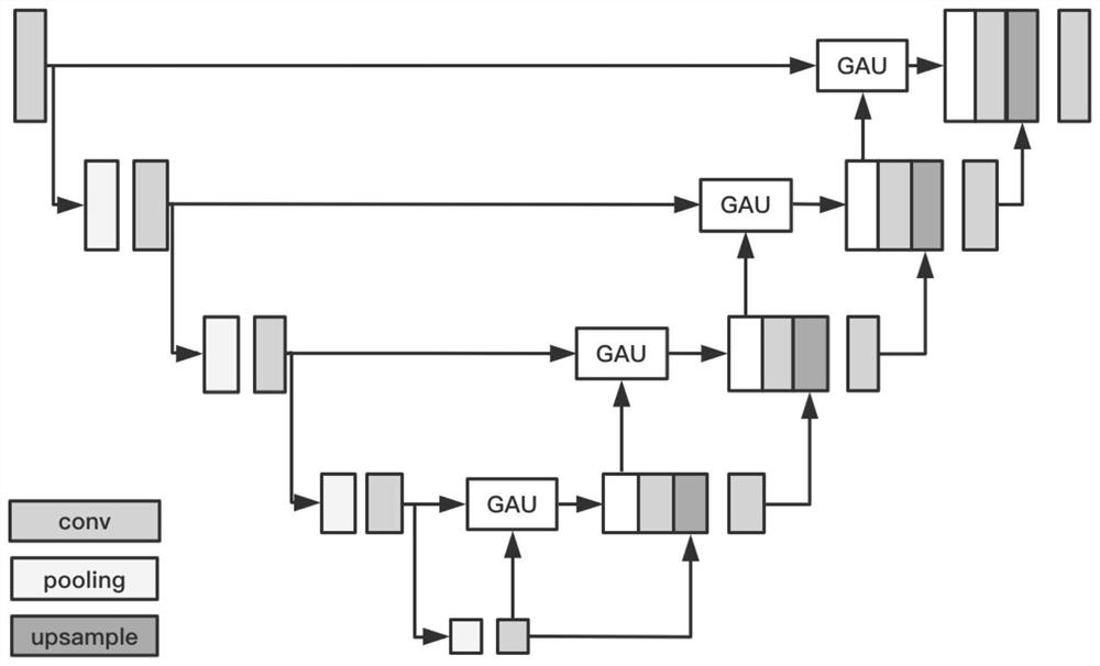
[0045] Step 2. Construct a liver organ segmentation network U based on global attention.

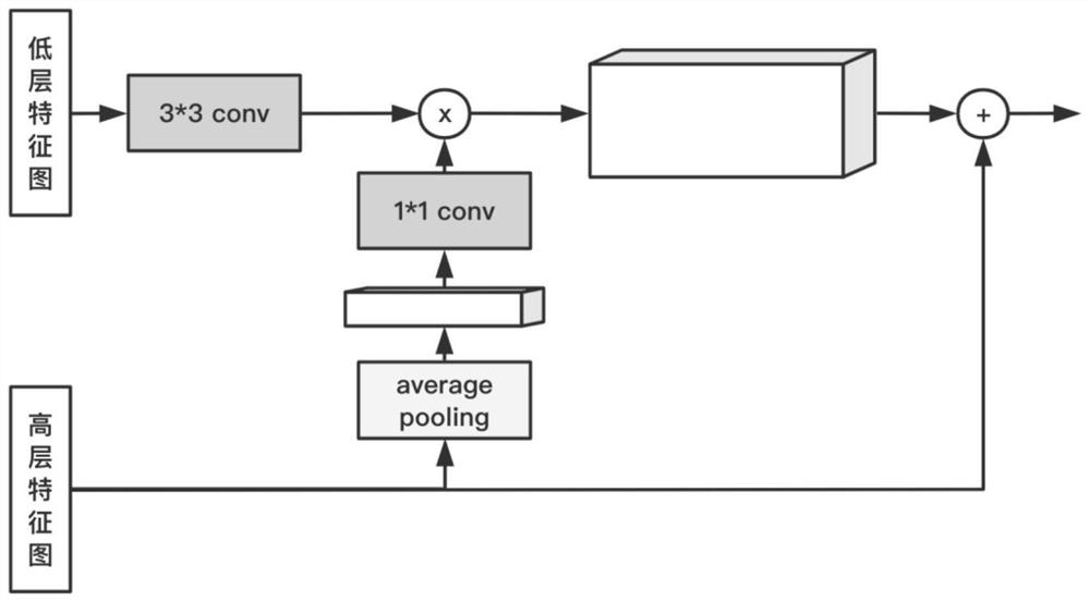
[0046] 2.1) Introduce the global attention upsampling GAU module in the existing segmentation network UNet:

[0047] The global attention upsampling GAU module, such as figure 2 As shown, it includes 3*3 convolutional layer, 1*1 convolutional layer, global pooling layer, input low-level feature map and high-level featu...


PUM


Abstract

The invention discloses a liver CT image multi-lesion classification method based on sample generation and transfer learning. The method mainly solves the problem that an existing method is not high in liver multi-lesion detection performance. The implementation scheme is as follows: dividing a data set; respectively constructing a liver organ segmentation network and a liver lesion detection network; based on the deep convolution generative adversarial network, constructing a liver cyst sample generation network and a liver hemangioma sample generation network, and respectively generating newliver cyst and liver hemangioma samples; constructing a liver lesion classification network; subjecting a liver CT image to be detected firstly to organ segmentation by using a liver organ segmentation network, then subjecting a segmentation result to lesion detection by using a liver lesion detection network, and finally classifying detected lesions by using a liver lesion classification network. According to the invention, imbalance of different types of sample sizes is relieved, the lesion classification performance is improved, and the method can be used for positioning and qualifying various lesions such as liver cancer, liver cyst and hepatic hemangioma existing in the liver CT image.

Description

technical field [0001] The invention belongs to the technical field of image processing, and further relates to a method for classifying liver lesions, which can be used to locate and characterize various lesions such as liver cancer, hepatic cysts, and hepatic hemangiomas in liver CT images. Background technique [0002] As the largest digestive gland in the human body, the liver plays an irreplaceable role. However, there are many kinds of diseases. For example, liver cancer is one of the common malignant tumors. The incidence rate is increasing year by year. The early manifestations are usually not obvious. Epigastric discomfort, pain, fever, and fatigue will only appear when the tumor is large or the course of the disease progresses to the middle and late stages. , nausea, loss of appetite and other symptoms. Imaging diagnosis is of great significance to the diagnosis and curative effect evaluation of liver cancer, liver cyst, hepatic hemangioma and other diseases. In ...

Claims


Application Information

Patent Timeline
IPC IPC(8): G06K9/62G06K9/34G06N3/04G06N3/08
CPCG06N3/084G06V10/267G06N3/045G06F18/2415Y02A90/10
Inventor 缑水平曹思颖周海彬杨玉林刘豪锋续溢男骆安琳
Owner XIDIAN UNIV
Why Eureka
Try Eureka