System and method for automatic segmentation, measurement and localization of breast masses on MRI

An automatic segmentation and mammary gland technology, applied in the field of medical information, can solve the problems of inability to automatically measure tumor size and tumor location information, reduce doctor's work efficiency, disadvantages, etc., achieve complete report content, save labeling time, and improve work efficiency.

Active Publication Date: 2022-07-15
北京赛迈特锐医疗科技有限公司
View PDF5 Cites 0 Cited by
  • Summary
  • Abstract
  • Description
  • Claims
  • Application Information

AI Technical Summary

Problems solved by technology

[0003] In view of this, the main purpose of the present invention is to provide a system and method for automatically segmenting, measuring, and locating breast masses on MRI, which can solve the problems in the prior art caused by the inability to automatically measure the mass size and mass location information. It is beneficial to guide the needle biopsy and reduce the work efficiency of doctors

Method used


Image

  • System and method for automatic segmentation, measurement and localization of breast masses on MRI
  • System and method for automatic segmentation, measurement and localization of breast masses on MRI
  • System and method for automatic segmentation, measurement and localization of breast masses on MRI

Examples


Embodiment 1

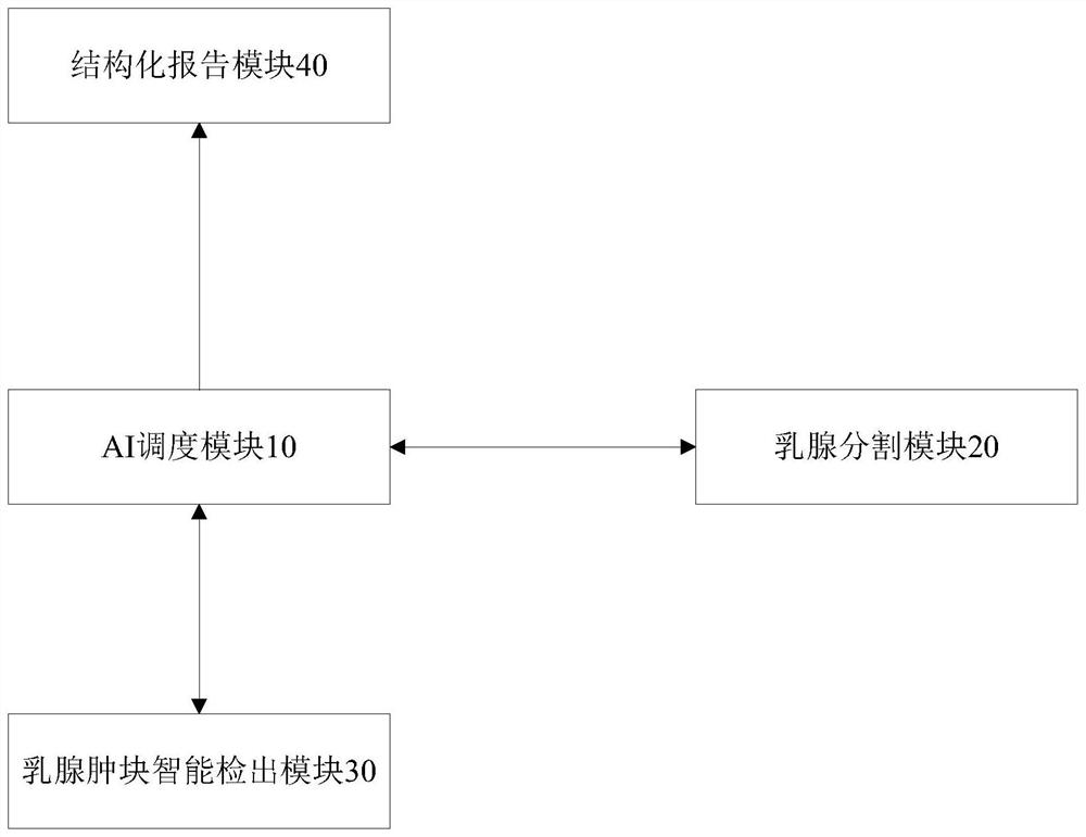
[0028] figure 1 A schematic structural diagram of a system for automatically segmenting, measuring and locating breast masses on MRI according to Embodiment 1 of the present invention is shown; as figure 1 As shown, the system includes: an AI scheduling module 10, a breast segmentation module 20, an intelligent breast mass detection module 30 and a structured reporting module 40, wherein,

[0029] The AI ​​scheduling module 10 is connected to the breast segmentation module 20, the breast mass intelligent detection module 30 and the structured reporting module 40 respectively, and is used for performing a breast dynamic enhanced MRI (DCE MRI) scan when the patient is scanned. During the inspection, extract the header file information of the DICOM image, search for the DCE sequence image based on the header file information, and send the DCE sequence image to the breast segmentation module 20 and the breast mass intelligent detection module 30;

[0030] When a patient has indic...

Embodiment 3

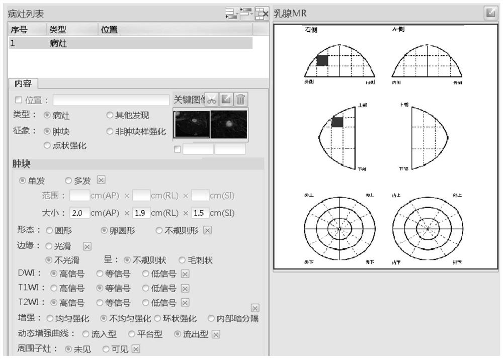
[0037] Figure 4 Shows the schematic diagrams of the partitions on the axial image, sagittal image, and clock face image of bilateral breast glands in the system for automatic segmentation, measurement and localization of breast masses on MRI according to Embodiment 3 of the present invention; like Figure 4 As shown, the rules for partitioning the axial images of bilateral breast glands are medial, lateral and anterior, middle, and posterior, and the axial images of bilateral breast glands are divided into 16 partitions; The sagittal images of breast glands are divided into upper, lower and anterior, middle, and posterior parts, and the sagittal images of bilateral breast glands are divided into 16 divisions; The division rules of the clock face image are center, middle, periphery, inner upper, inner lower, outer upper, outer lower, and each is divided into 36 divisions on the clock face image of bilateral breast glands.

[0038] Partitions 1-16 are axial images of the righ...

Embodiment 4

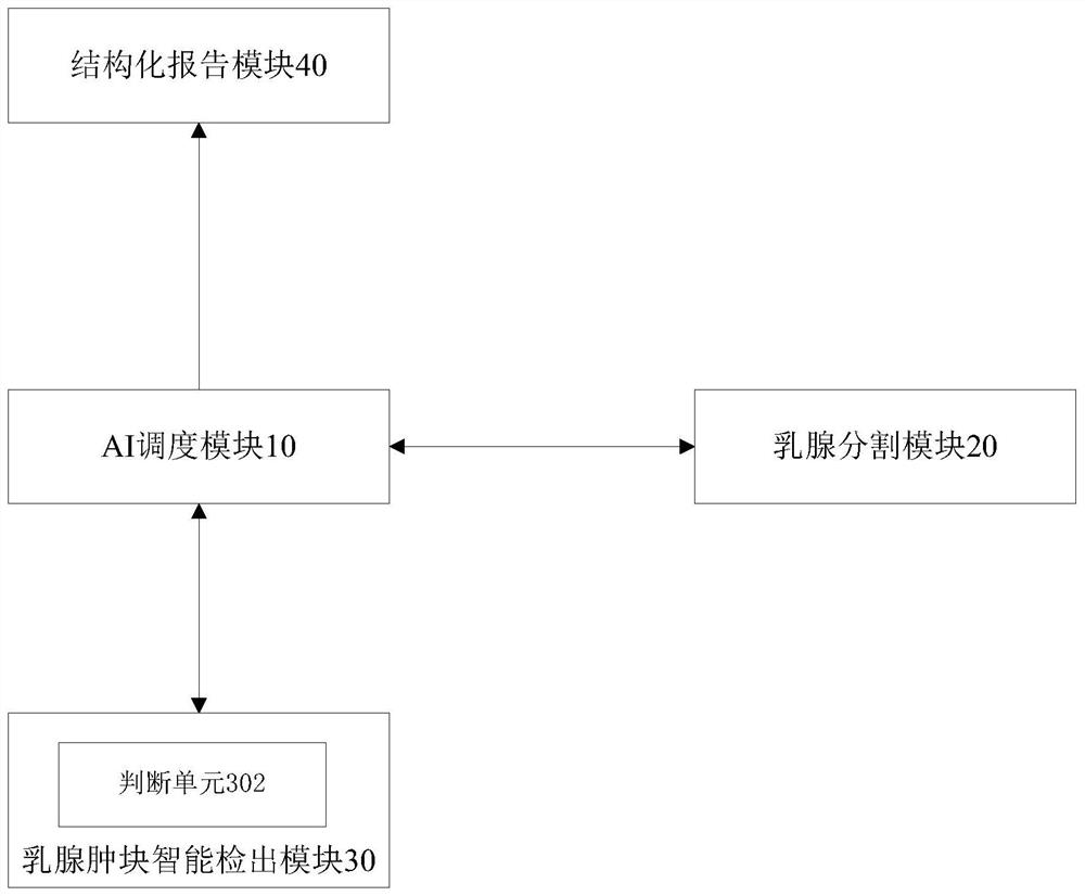
[0046] Figure 5 A schematic structural diagram of a system for automatically segmenting, measuring and locating breast masses on MRI according to Embodiment 4 of the present invention is shown; as Figure 5 As shown, the structured reporting module 40 further includes a navigation map generating unit 402, which is connected to the AI ​​scheduling module 10, and is used to receive all cancer foci data whose shortest diameter is greater than or equal to a preset threshold, and automatically mark the location of the cancer foci on the navigation map. , and display the marked navigation graph in the corresponding position of the structured report interface.

[0047] The embodiment of the present invention is provided with a navigation map generation unit, which can receive all the cancer foci data whose shortest diameter is greater than or equal to a preset threshold, automatically mark the location of the cancer foci on the navigation map, and display the marked navigation map i...


PUM


Abstract

The invention provides a system for automatically segmenting, measuring and locating breast masses on MRI, including an AI scheduling module to extract header file information of a patient's DICOM image and search for a DCE sequence; a breast segmentation module to segment the bilateral breast glands of the DCE sequence , on the axial image, sagittal image and clock plane image of bilateral breast glands, respectively, divide the bilateral breast glands into multiple different partitions, and set a unique partition number for each partition; The intelligent detection module identifies all breast cancer foci in the DCE sequence, measures the three-dimensional diameter and volume of the cancer foci, and matches any two partition data of the three partition data of the axial, sagittal, and clock plane images. The location of cancer foci can be output; the structured report module outputs the volume, number, and location of cancer foci. The invention also discloses a method for automatically segmenting, measuring and locating breast masses on MRI. The invention can accurately locate the position and size of the cancer foci and improve the working efficiency of doctors.

Description

technical field [0001] The present invention relates to the field of medical information, and more particularly, to a system and method for automatically segmenting, measuring and locating breast masses on MRI. Background technique [0002] The main tasks of breast magnetic resonance imaging (Magnetic Resonance Imaging, MRI) are: 1) Detect the mass, measure the size and location, determine whether breast-conserving surgery is feasible, and provide a basis for formulating a surgical plan. There are various treatment methods for breast cancer, and breast-conserving surgery and neoadjuvant chemotherapy are the main treatment methods. Before breast-conserving surgery, it is necessary to know whether the mass is single or multiple, whether it is located in one or both breasts, occupying several quadrants, and its relationship with the pectoralis major muscle to determine whether breast-conserving surgery is feasible. 2) Lesion screening in high-risk breast cancer patients with s...

Claims


Application Information

Patent Timeline
Patent Type & Authority Patents(China)
IPC IPC(8): A61B5/055
CPCA61B5/055A61B5/4312
Inventor 岳新贺长征张虽虽王霄英
Owner 北京赛迈特锐医疗科技有限公司
Why Eureka
Try Eureka