
Automatic segmentation method for oral cancer epithelial tissue region of pathology image

A technology of epithelial tissue and pathology, applied in the fields of machine learning and medical image processing, can solve the problems of difficult aggregation of staining variability information, time-consuming and boring, etc.

Pending Publication Date: 2021-04-27
SHAANXI NORMAL UNIV +1
View PDF4 Cites 6 Cited by
  • Summary
  • Abstract
  • Description
  • Claims
  • Application Information

AI Technical Summary

Problems solved by technology

However, when applying ML / DL to more specific regions, in WSI epithelial segmentation, it may encounter time-consuming and boring issues such as GroundTruth annotation, variability of staining, and difficulty in processing image information based on patches. gathering challenge

Method used


Image

  • Automatic segmentation method for oral cancer epithelial tissue region of pathology image
  • Automatic segmentation method for oral cancer epithelial tissue region of pathology image
  • Automatic segmentation method for oral cancer epithelial tissue region of pathology image

Examples


Embodiment Construction

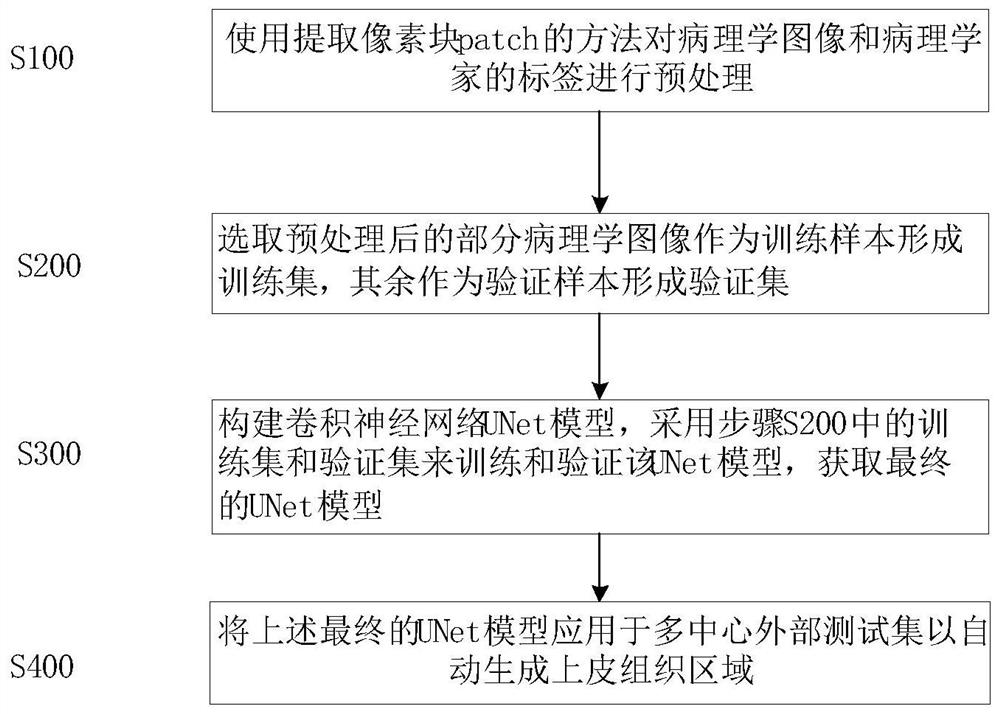
[0019] In one embodiment, such as figure 1 As shown, a method for automatic segmentation of oral cancer epithelial tissue regions that provides a pathological image is disclosed, which includes the following steps:

[0020] S100: Preprocessing the pathology image and the pathologist's label by using a method of extracting a pixel block patch;

[0021] S200: selecting part of the preprocessed pathological images as training samples to form a training set, and the rest as verification samples to form a verification set;

[0022] S300: Construct a convolutional neural network UNet model, use the training set and verification set in step S200 to train and verify the UNet model, and obtain the final UNet model;

[0023] S400: Apply the above final UNet model to a multi-center external test set to automatically generate epithelial tissue regions.

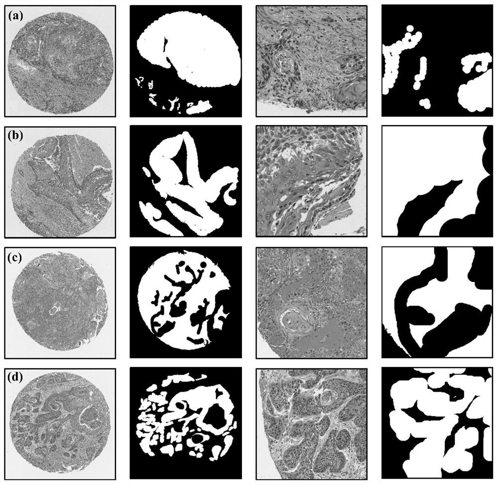
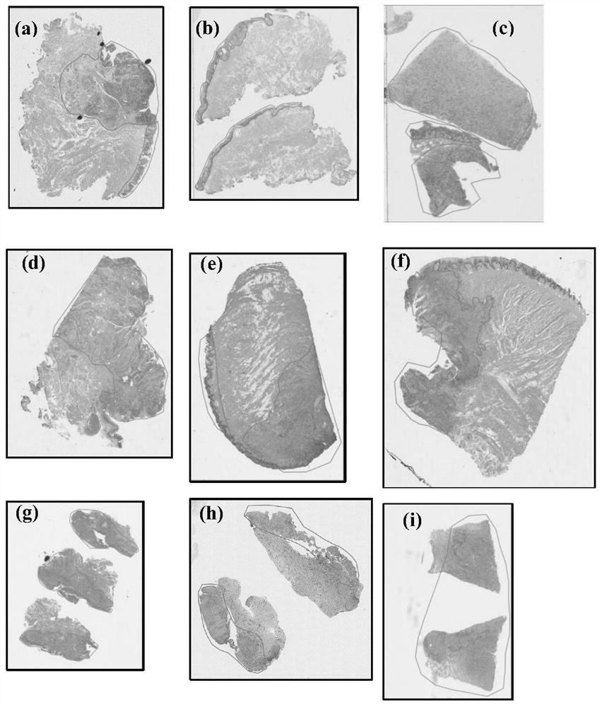
[0024] For this example, a UNet-based deep learning framework was used to segment epithelial regions from two types of histopathology ...


PUM


Abstract

The invention discloses an automatic segmentation method for an oral cancer epithelial tissue region of a pathological image. The method comprises the following steps: S100, preprocessing the pathological image and a label of a pathologist by using a method for extracting a pixel block patch; s200, selecting part of the preprocessed pathological images as training samples to form a training set, and taking the rest pathological images as verification samples to form a verification set; s300, constructing a convolutional neural network UNet model, and training and verifying the UNet model by adopting the training set and the verification set in the step S200 to obtain a final UNet model; and S400, applying the final UNet model to a multi-center external test set to automatically generate an epithelial tissue region. According to the method, training, verification and testing are not carried out on each data set in head and neck cancer detection, only TMA data is used for training, testing is carried out on a multi-center external WSI data set, and the method is more persuasive to unknown images.

Description

technical field [0001] The disclosure belongs to the technical fields of medical image processing and machine learning, and in particular relates to a method for automatically segmenting oral cancer epithelial tissue regions of pathological images. Background technique [0002] Oral squamous cell carcinoma (OC-SCC) is the most common malignancy of the head and neck worldwide, with high invasiveness to adjacent tissues and metastatic potential to distant organs. Better diagnostic and risk stratification tools are therefore needed for personalized treatment of OC-SCC patients. [0003] The pathologist's approach to diagnosis and risk factor stratification begins with the identification of the tumor region. These areas are usually analyzed by a pathologist by light microscopy using routinely stained tissue. The entire tumor area is composed of cancer cells and their supporting tissue matrix. In the case of carcinoma, the epithelial tumor area is the most evaluated part. His...

Claims


Application Information

Patent Timeline
Patent Type & Authority Applications(China)
IPC IPC(8): G06T7/00G06T7/11G06N3/04G06N3/08
CPCG06T7/0012G06T7/11G06N3/08G06T2207/30096G06N3/048G06N3/045
Inventor 陆铖吴玉欣王亮郭玉川
Owner SHAANXI NORMAL UNIV
Learn More