Method of lifting an epithelial layer and placing a corrective lens beneath it

a technology of epithelial layer and corrective lens, which is applied in the field of living lenses, can solve the problems of spherical ammetropia, formation of image, and eye contact with ophthalmologist or optometris

Inactive Publication Date: 2005-04-19
TISSUE ENG REFRACTION
View PDF23 Cites 48 Cited by
  • Summary
  • Abstract
  • Description
  • Claims
  • Application Information

AI Technical Summary

Problems solved by technology

The most common problem an ophthalmologist or optometrist will encounter is that of spherical ammetropia, or the formation of an image by the eye which is out of focus with accommodation due to an improperly shaped globe.
These techniques and their variations were generally considered to be unsuccessful due to frequent complications involving irregular astigmatism, delayed surgical healing, corneal scarring, and instability of the refractive result.
The problems were attributed to the technical complexity of the procedures as well as to the distortion in architecture of the corneal tissue secondary to lens manipulation.
Freezing of the lenticule in keratophakia and keratomileusis also causes severe damage to epithelial and stromal cells and disrupts the lamellar architecture of the cornea.
However, this lens is frozen prior to reshaping on a lathe which results in stromal keratocyte death.
Furthermore, the cited documents do not show any methods of lens preparation that remove native corneal tissue cells and replace them with cells cultivated from human cornea.
The tissue using these cell culturing procedures is also quite fragile.
However, the laser changes the native structure of the cornea by irreversibly coagulating collagen.
Post-laser relaxation of collagen is not possible with this treatment.
The inventive corneal overlay technique may be completed under local anesthesia as well as general anesthesia, and the availability of a precut lens will greatly decrease procedure time, patient cost, and risk of operative complications.

Method used


Image

  • Method of lifting an epithelial layer and placing a corrective lens beneath it
  • Method of lifting an epithelial layer and placing a corrective lens beneath it
  • Method of lifting an epithelial layer and placing a corrective lens beneath it

Examples


Embodiment Construction

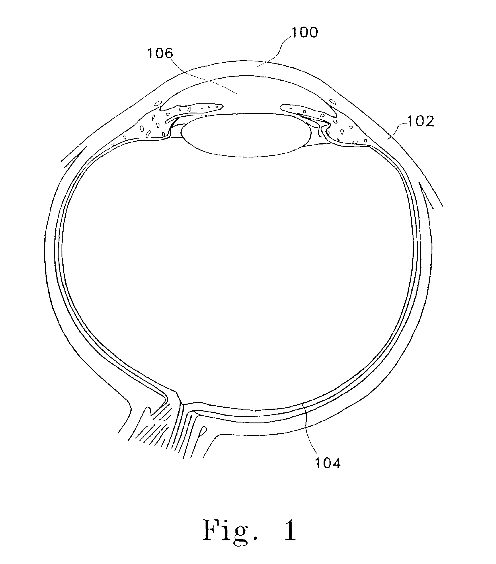
[0037]The eye is designed to focus light onto specialized receptors in the retina that turn quanta of light energy into nerve action potentials. As shown in FIG. 1, light rays are first transmitted through the cornea (100) of the eye. The cornea is transparent due to the highly organized structure of its collagen fibrils. The margins of the cornea merge with a tough fibrocollagenous sclera (102) and is referred to as the corneo-scleral layer.

[0038]The cornea (100) is the portion of the corneo-scleral layer enclosing the anterior one-sixth of the eye. The smooth curvature of the cornea is the major focusing power of images on the retina (104) and it provides much of the eye's 60 diopters of converging power. The cornea is an avascular structure and is sustained by diffusion of nutrients and oxygen from the aqueous humor (106). Some oxygen is also derived from the external environment. The avascular nature of the cornea decreases the immunogenicity of the tissue, increasing the succes...


PUM

PropertyMeasurementUnit
diameteraaaaaaaaaa
diameteraaaaaaaaaa
diameteraaaaaaaaaa

Abstract

This relates to a lens made of donor corneal tissue suitable for use as a contact lens or an implanted lens, to a method of preparing that lens, and to a technique of placing the lens on the eye. The lens is made of donor corneal tissue that is acellularized by removing native epithelium and keratocytes. These cells optionally are replaced with human epithelium and keratocytes to form a lens that has a structural anatomy similar to human cornea. The ocular lens may be used to correct conditions such as astigmatism, myopia, aphakia, and presbyopia.

Description

RELATED APPLICATIONS[0001]This is a continuation of PCT Application No. PCT / US01 / 22633, having an International Filing Date of Jul. 18, 2001 and a continuation-in-part of U.S. patent application Ser. No. 09 / 618,580, filed Jul. 18, 2000 now U.S. Pat No. 6,544,286.FIELD OF THE INVENTION[0002]This invention is in the field of ophthalmology. More particularly, it relates to a living lens, suitable for use as a contact lens or for subepithelial implantation. The lens is made of donor corneal tissue. The invention includes methods of preparing that lens and to techniques of placing the lens on the eye.BACKGROUND OF THE INVENTION[0003]The visual system allows the eye to focus light rays into meaningful images. The most common problem an ophthalmologist or optometrist will encounter is that of spherical ammetropia, or the formation of an image by the eye which is out of focus with accommodation due to an improperly shaped globe. The ophthalmologist or optometrist determines the refractive s...

Claims


Application Information

Patent Timeline
Patent Type & Authority Patents(United States)
IPC IPC(8): A61L27/00A61L27/36A61F2/14G02C7/04A61F2/16A61F9/007
CPCA61F2/142A61F2/145A61L27/3604A61L27/3683A61L27/3813A61L27/3839A61L27/3641A61L2430/16A61L27/36
Inventor PEREZ, EDWARD
Owner TISSUE ENG REFRACTION
Why Eureka
Try Eureka