Protein-free defined media for the growth of normal human keratinocytes

a keratinocyte and protein-free technology, applied in the field of biochemistry, can solve the problems of terminal differentiation, inability to control, and inability to grow achieve the effects of reducing the cell division capacity of normal human epithelial stem cells, and reducing the cell division capacity

Inactive Publication Date: 2006-05-02
BIOPLAST MEDICAL
View PDF2 Cites 46 Cited by
  • Summary
  • Abstract
  • Description
  • Claims
  • Application Information

AI Technical Summary

Benefits of technology

[0041]In a more preferred embodiment of the cell competency solution, the sodium chloride is at a concentration of 100 to 130 mM; the HEPES is at a concentration of 18 to 21 mM; the trypsin is at a concentration of 0.12 to 0. 18% w / v in the digestion solution and the soybean trypsin inhibitor is at a concentration of 0.3 to 0.8% w / v in the second CCS.

Problems solved by technology

In contrast, the culture of normal human epithelial stem cells has presented many difficulties not the least of which is the inexorable tendency for these cells to undergo uncontrolled, irreversible, terminal differentiation with the consequent loss of cell division capacity.
However, the actual composition of the SFM media is not disclosed other than it does not contain the growth promoting additives insulin, epidermal growth factor and fibroblast growth factor.
Lower concentration of all-trans retinoic acid were ineffective.
This procedure has serious limitations for large scale production of human epithelium as the use of serum inextricably confounds the culture of purely basal cells with the dynamics of serum-induced differentiation.
Moreover, the process of Green et al. does not describe the formation of a histologically complete epidermis.
The use of any organotypic substrate as well as feeder or companion cell types, e.g. fibroblasts, seriously limits the resulting products safety and economic viability.
The prior art media which contain undefined serum and / or feeder cell factors and / or organotypic substrates and millimolar concentrations of Ca2+, high levels of buffers, inadequate levels of amino acids and incorrect osmolalities were not designed for the unlimited proliferation of undifferentiated basal cells.

Method used


Image

  • Protein-free defined media for the growth of normal human keratinocytes
  • Protein-free defined media for the growth of normal human keratinocytes
  • Protein-free defined media for the growth of normal human keratinocytes

Examples


example 1

Primary and Secondary Culture of Normal Human Epidermal Keratinocytes in HECK-110 Protein-Free Defined Medium

Isolation of Basal Cells and Primary Cultures

[0119]Primary cultures of normal human basal epidermal keratinocytes were started by subjecting full-thickness skin samples to enzymatic digestion. Skin obtained from biopsies or autopsies was first cleaned of adhering subdermal fat and the dermis was reduced to less than 3 mm in thickness. The skin sample was then cut into 8 to 12 small pieces (usually 0.5 cm2). These pieces were floated on top of sterile CCS (Cell Competency Solution). CCS consisted of glucose, 10 mM; KCl, 3 mM; NaCl, 130 mM; Na2HPO4.7H2O, 1 mM; phenol red, 3.3 μM; HEPES at 23 mM; (See Shipley, G. D. and Ham, R. G., In Vitro 17:656-670 (1981)) and 0.17% trypsin (w / v) and 100 units / ml of both penicillin and streptomycin. After 14 to 16 hours of digestion at 4° C., the dermis was separated from the epidermis by a split-dermis technique. This was accomplished by pla...

example 2

Preparation of HECK-110 Basal Nutrient Medium

[0124]One aspect of the present invention relates to the preparation of a new media suitable for the large scale amplification of both primary and secondary cultures of normal human epithelial cells, such as keratinocytes, and for conversion of proliferating normal human epithelial monolayer cultures to a fully differentiated tissue transplantable to a human being. More particularly, this Example 2 is directed to the materials and procedures for preparation of a basal nutrient medium (Human Epidermal Cell Keratinocyte, HECK-110), and experiments evidencing its superiority in stimulating epithelial cell growth. The media according to this invention are novel and unobvious by design of the osmolarity, toxicity and pH-buffering properties.

[0125]Table 1 below details the concentration of components in basal medium, HECK-110. All biochemicals, growth factors and hormones were purchased from Sigma Chemical Company (St. Louis, Mo., U.S.A.), and ...

example 3

Growth of Secondary Cultures of Normal Human Epidermal Keratincytes in HECK-110 Protein-free Defined Medium

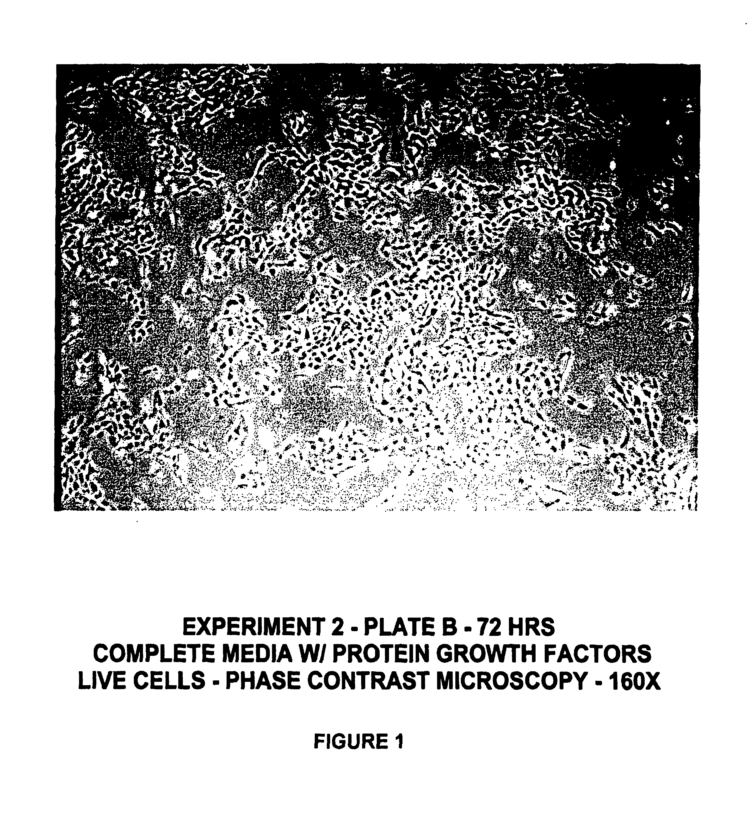
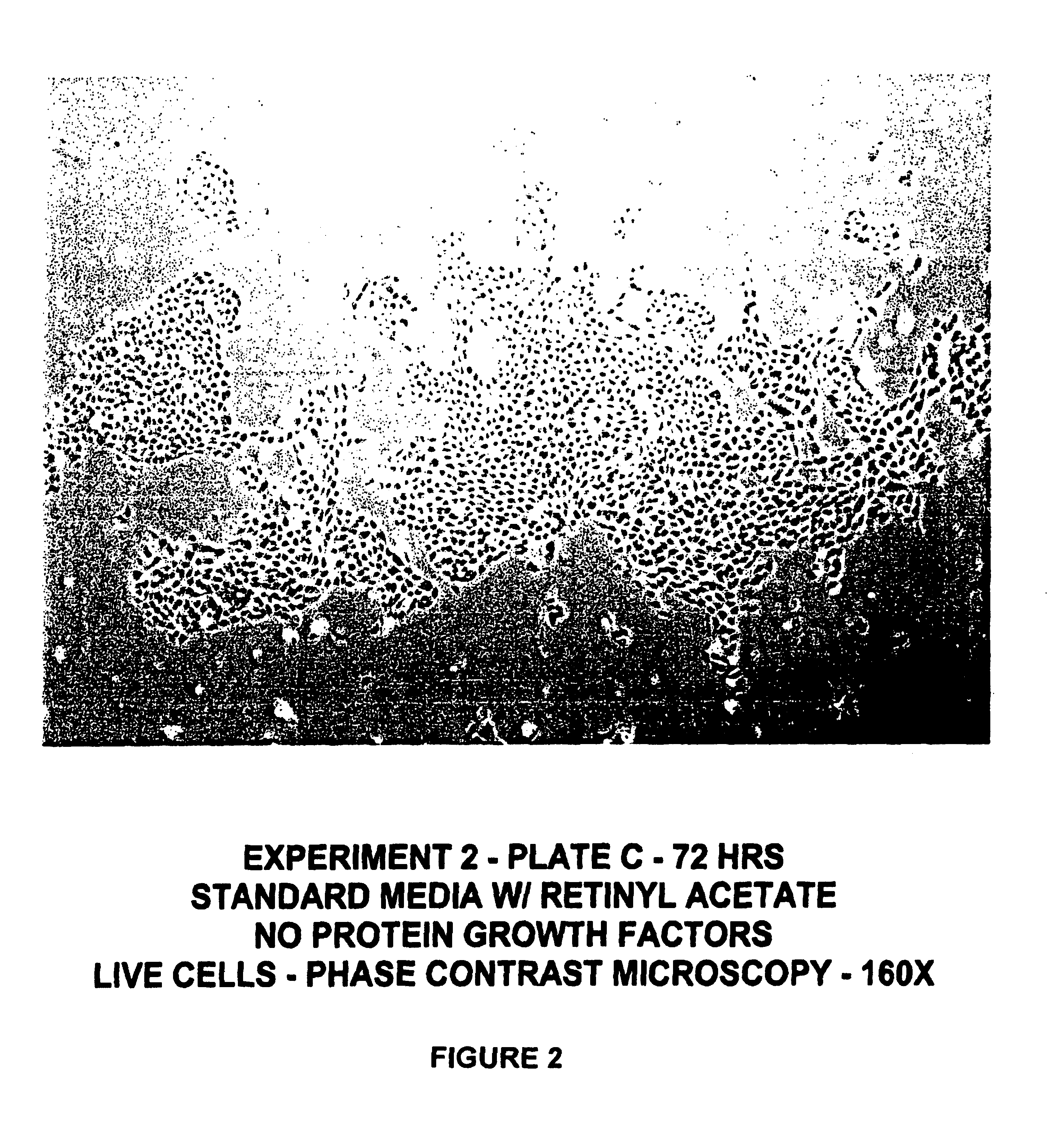
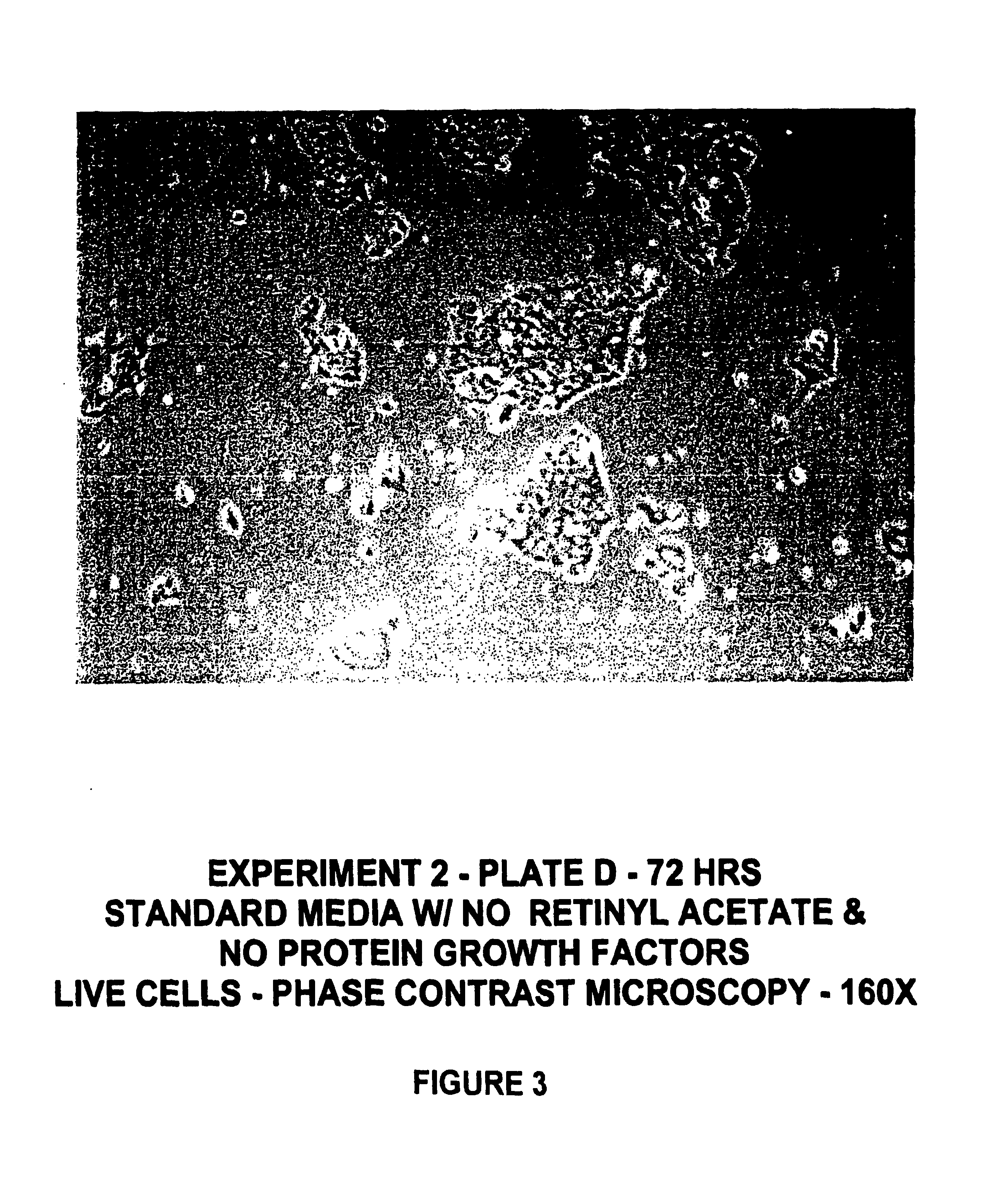
[0131]Human keratinocyte cultures were initiated from neonatal foreskin as described in Example 1, and then placed in secondary culture in complete HECK-109 FS medium. The purpose of the following experiment was to determine the effect of retinyl acetate on the proliferation of keratinocyte cultures refed HECK-109 basal medium lacking EGF and IGF-1 and supplemented with hydrocortisone, ethanolamine, and phosphoethanolamine. For this purpose, duplicate secondary cultures were refed either 1) complete HECK-109 FS medium (positive control) containing 0.1 mM Ca2+, 2) standard HECK-109 medium, i.e., basal medium supplemented with only hydrocortisone, ethanolamine, and phosphoethanolamine, and containing 0.1 mM Ca2+ and 3) standard HECK-109 supplemented with retinyl acetate (3×10⁻⁸M) (now called HECK-110). FIGS. 1-3 are phase contrast microscope photomicrographs of living cultures of...


PUM

PropertyMeasurementUnit
concentrationaaaaaaaaaa

Abstract

Improvements are made to a novel media that replace the requirement for all protein growth factors by the addition to the medium of physiological concentrations of retinyl acetate. The media are serum-free, companion cell or feeder layer-free and organotypic, matrix free solutions for the cultivation of clonally competent basal keratinocytes. The media and methods are useful in the production of epidermal epithelial tissue that is suitable for skin grafting.

Description

RELATED APPLICATIONS[0001]This application is a continuation-in-part of U.S. application Ser. No. 09 / 271,777, now U.S. Pat. No. 6,162,643, issued Dec. 19, 2000, entitled PROCESS AND MEDIA FOR THE GROWTH OF HUMAN EPITHELIA; which is a continuation of U.S. application Ser. No. 09 / 133,386, filed Aug. 13, 1998, now U.S. Pat. No. 5,912,175; which is a continuation of U.S. application Ser. No. 08 / 893,195, filed Jul. 15, 1997, now U.S. Pat. No. 5,834,312 issued Nov. 10, 1998, entitled PROCESS AND MEDIA FOR THE GROWTH OF HUMAN EPITHELIA; which is a continuation-in-part of U.S. application Ser. No. 08 / 500,744, filed Jul. 11, 1995, now U.S. Pat. No. 5,686,307 issued Nov. 11, 1997, entitled SERUM-FREE MEDIUM FOR USE IN THE FORMATION OF A HISTOLOGICALLY COMPLETE LIVING HUMAN SKIN SUBSTITUTE; which is a continuation of U.S. application Ser. No. 08 / 318,221, filed Oct. 5, 1994, now abandoned; which is a continuation of U.S. application Ser. No. 08 / 184,905, filed Jan. 21, 1994, now abandoned; which...

Claims


Application Information

Patent Timeline
Patent Type & Authority Patents(United States)
IPC IPC(8): C12N5/00
CPCC12N5/0629C12N2500/20C12N2500/32C12N2500/36C12N2501/15C12N2500/99C12N2501/105C12N2501/11C12N2500/60C12N2500/90C12N2500/95
Inventor WILLE, JR., JOHN J.
Owner BIOPLAST MEDICAL
Why Eureka
Try Eureka