Picture screening method and system for esophageal cancer model training and storage medium

A screening method and model training technology, applied in the field of image screening methods, systems and storage media, can solve problems such as poor model generalization ability, surge in model calculations, and increased data volume

Pending Publication Date: 2021-06-11
成都微识医疗设备有限公司
View PDF0 Cites 3 Cited by
  • Summary
  • Abstract
  • Description
  • Claims
  • Application Information

AI Technical Summary

Problems solved by technology

However, for the sample data to completely cover the sample space, the amount of data required will increase exponentially
The large-sample training model not only increases the amount of calculations, but also has a large difference in the amount of data of different categories. If the balance of samples is

Method used


Image

  • Picture screening method and system for esophageal cancer model training and storage medium
  • Picture screening method and system for esophageal cancer model training and storage medium
  • Picture screening method and system for esophageal cancer model training and storage medium

Examples


Embodiment 1

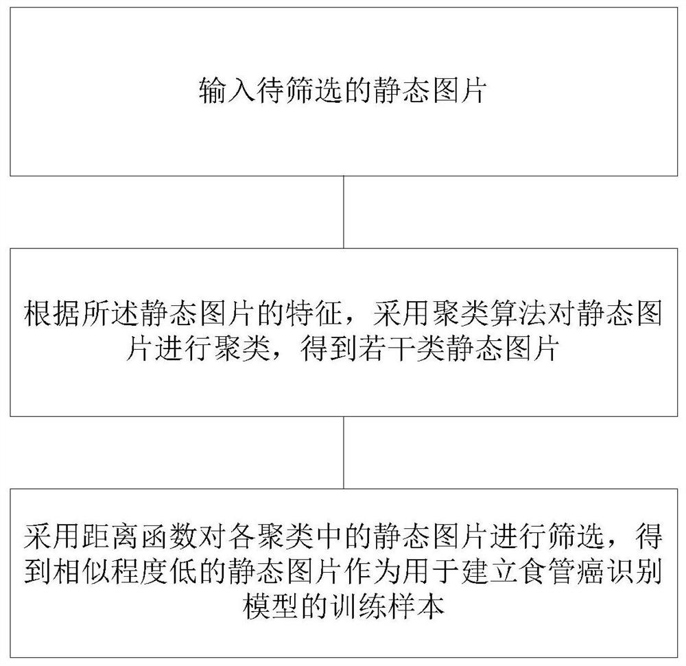
[0050] like figure 1 The shown screening method for pictures used for esophageal cancer model training comprises the following steps:

[0051] Enter the static picture to be filtered;

[0052] According to the feature of described static picture, adopt clustering algorithm to carry out clustering to static picture, obtain several classes of static pictures;

[0053] The distance function was used to screen the static pictures in each cluster, and the static pictures with low similarity were obtained as training samples for establishing an esophageal cancer recognition model.

[0054] The step of using the distance function to filter the static pictures in each cluster includes:

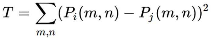
[0055] A distance function is used to calculate the distance values ​​between all still pictures in the same cluster, and the distance function is:

[0056]

[0057] Among them, P i and P j are respectively the i-th static picture and the j-th static picture in the same cluster, P i (m,n) is ...

Embodiment 2

[0067] A screening system for images used in esophageal cancer model training, including:

[0068] The input module is used for inputting static pictures to be screened;

[0069] The screening module is used to apply a clustering algorithm to cluster the static pictures to obtain several categories of static pictures, and use a distance function to screen the static pictures in each cluster to obtain static pictures with a low degree of similarity as used for establishing an esophageal cancer identification model training samples;

[0070] The output module outputs the training samples obtained by screening;

[0071] The steps of the screening module using a distance function to screen the static pictures in each cluster include:

[0072] A distance function is used to calculate the distance values ​​between all still pictures in the same cluster, and the distance function is:

[0073]

[0074] Among them, P i and P j are respectively the i-th static picture and the j-...

Embodiment 3

[0080] A storage medium, including a stored computer program, wherein, when the computer program is running, the device where the storage medium is located is controlled to execute the screening method described in any of the foregoing embodiments.

[0081] In this embodiment, the above-mentioned storage medium is a computer-readable storage medium. If the colonoscopy quality assessment device is implemented in the form of a software function unit and sold or used as an independent product, it can be stored in a computer-readable storage medium. medium. Based on this understanding, the present invention realizes all or part of the processes in the methods of the above embodiments, and can also be completed by instructing related hardware through computer programs. The computer program can be stored in a computer-readable storage medium, and the computer When the program is executed by the processor, the steps in the above-mentioned various method embodiments can be realized. ...


PUM


Abstract

The invention discloses a picture screening method and system for esophageal cancer model training, and a storage medium. The method comprises the following steps: inputting a to-be-screened static picture; clustering the static pictures by adopting a clustering algorithm according to the characteristics of the static pictures to obtain a plurality of types of static pictures; and screening the static pictures in each cluster by adopting a distance function to obtain static pictures with low similarity as training samples for establishing an esophageal cancer recognition model. According to the method, when the static pictures are input, a larger sample size can be allowed to be adopted to solve the problem of poor model generalization ability, meanwhile, the static pictures with large samples are clustered through a clustering algorithm, then the static pictures with low similarity in each cluster are screened through a distance function, and the static pictures with high similarity are obtained. And finally, on the premise that the sample coverage rate is not obviously influenced, the transformation from a large sample to a small sample is realized, and an esophageal squamous carcinoma lesion picture suitable for training and identifying narrow-band imaging is obtained.

Description

technical field [0001] The invention relates to the field of intelligent medical technology, in particular to a method, system and storage medium for screening images used for esophageal cancer model training. Background technique [0002] Esophageal cancer is one of the most common malignant tumors of the digestive tract in the world, and its pathological types mainly include esophageal squamous cell carcinoma (ESCC) and esophageal adenocarcinoma (EADC). Upper gastrointestinal endoscopy combined with histopathology is the gold standard for the diagnosis of esophageal squamous cell carcinoma. For difficult-to-find lesions, chromoendoscopy and electronic chromoendoscopy are mainly used to find them, and then targeted biopsy for diagnosis through histopathology. [0003] With the development of image recognition technology, by screening and classifying lesion pictures, training and deep learning the characteristics of lesion and non-lesion pictures to establish a deep learnin...

Claims


Application Information

Patent Timeline
IPC IPC(8): G06T7/00G06K9/62G16H50/20
CPCG06T7/0012G16H50/20G06F18/2321G06F18/214
Inventor 肖潇刘敬家
Owner 成都微识医疗设备有限公司
Why Eureka
Try Eureka