CTP image-based infarction false positive filtering method

A filtering and image technology, which is applied in the field of medical image processing, can solve the problems that affect the diagnostic accuracy and false positives, and achieve the effect of improving accuracy and reducing the probability of false positives

Pending Publication Date: 2021-06-25
数坤(北京)网络科技股份有限公司
View PDF0 Cites 1 Cited by
  • Summary
  • Abstract
  • Description
  • Claims
  • Application Information

AI Technical Summary

Problems solved by technology

Automated image processing of CTP images to determine the location of the infarct nucleus can provide doctors with a basis for diagnosis and can effectively save time in the emergency procedure. Part of the ventricle or brain sulcus area is regarded as the infarct nucleus, resulting in false positives and affecting the accuracy of diagnosis

Method used


Image

  • CTP image-based infarction false positive filtering method

Examples


Embodiment

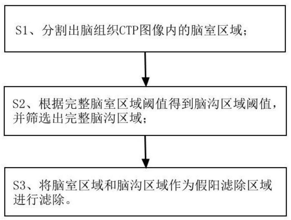
[0029] Such as figure 1 Shown, a kind of infarct false positive filtering method based on CTP image comprises the following steps:

[0030] S1. Segment the complete ventricle area in the brain tissue CTP image;

[0031] The step S1 is specifically implemented through the following steps:

[0032] S11. Input the CTP image of the brain tissue, and use the convolutional neural network to segment the preliminary ventricle area; the convolutional neural network adopts 3D-Unet.

[0033] S12. Calculate the mean CT value of the preliminary ventricle area as a threshold;

[0034] S13. Perform expansion processing on the preliminary ventricle area to obtain an expanded area, and use an area of ​​the expanded area lower than the threshold as a ventricle;

[0035] S14. Calculate the outer ventricular contour according to the threshold, and fill the non-preliminary ventricular region in the outer ventricular contour to obtain a complete ventricular region.

[0036] The calculation of t...


PUM


Abstract

The invention discloses a CTP image-based infarction false positive filtering method. The infarct false positive filtering method comprises the following steps: S1, segmenting a complete ventricle area in a brain tissue CTP image; S2, obtaining a brain sulcus area threshold according to the complete ventricle area threshold, and screening out a complete brain sulcus area; and S3, filtering the ventricle area and the cerebral sulcus area as false positive filtering areas. The complete ventricular region in the CTP image is firstly segmented, then the threshold value of the cerebral sulcus area is obtained according to the threshold value of the ventricle area, then the complete cerebral sulcus area is screened out, and the ventricular region and the cerebral sulcus area are used as false positive filtering regions for filtering, so that false positive probability is reduced, and diagnosis accuracy is improved.

Description

technical field [0001] The invention relates to the technical field of medical image processing, in particular to a method for filtering out infarction and false positives based on CTP images. Background technique [0002] As a very important imaging tool in the diagnosis and treatment of stroke, CTP cerebral perfusion imaging can accurately reflect the cerebral blood perfusion and is an effective means for early diagnosis of cerebral infarction, guidance of emergency thrombolytic therapy and prognosis. Automated image processing of CTP images to determine the location of the infarct nucleus can provide doctors with a basis for diagnosis and can effectively save time in the emergency procedure. As a result, part of the ventricle or brain sulcus area is regarded as the infarct nucleus, resulting in false positives and affecting the accuracy of diagnosis. Contents of the invention [0003] In order to solve the above problems, the present invention provides a method for fil...

Claims


Application Information

Patent Timeline
IPC IPC(8): G06T7/11G06T5/00G06T5/30G06T7/13G06T7/187G06T7/136G06N3/04
CPCG06T7/11G06T5/30G06T7/13G06T7/187G06T7/136G06T2207/10081G06T2207/30016G06N3/045G06T5/94
Inventor 左伟阳光张宇
Owner 数坤(北京)网络科技股份有限公司
Why Eureka
Try Eureka