Gynecological examination device

A technology for gynecological examination and inspection team, applied in the field of gynecological examiners, can solve the problems of low screening efficiency, long inspection duration, low one-time diagnosis efficiency, etc., and achieve the effect of high screening efficiency and short duration

Active Publication Date: 2021-07-13
SUNLAND MEDICAL CO LTD
View PDF6 Cites 6 Cited by
  • Summary
  • Abstract
  • Description
  • Claims
  • Application Information

AI Technical Summary

Problems solved by technology

[0003] In order to improve the screening efficiency of gynecological diseases, medical device products for screening are constantly being innovated and upgraded at home and abroad. The light source and lens are integrated into the vaginal dilator to form a visual module, which can improve the efficiency of preliminary inspection. However, this kind of gynecological medical treatment The improvement of the efficiency of the preliminary inspection of the device is aimed at the situation that there is no abnormality in the inspection. When the abnormality is found in the preliminary inspection, the doctor needs to replace the colposcope for further verification and confirmation, resulting in a longer overall inspection time, low one-time diagnosis efficiency, and overall screening Check efficiency is low

Method used


Image

  • Gynecological examination device
  • Gynecological examination device
  • Gynecological examination device

Examples


Embodiment 1

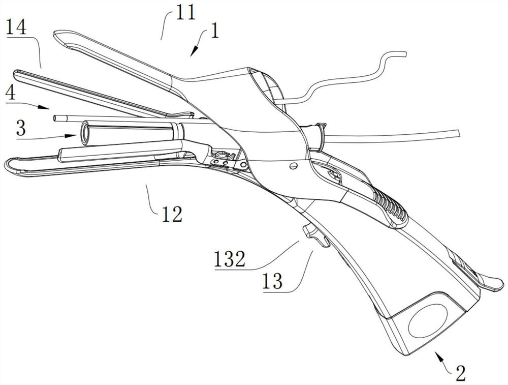
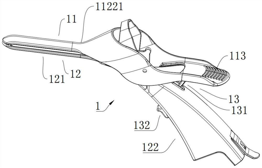
[0052] refer to figure 1 with figure 2 , figure 1 It is the structural representation of the gynecological examination device in the embodiment, figure 2 It is a structural schematic diagram of the expansion mechanism in the closed state in the embodiment. The gynecological examination device in this embodiment includes an expansion mechanism 1 , a visual mechanism 2 and an inspection mechanism 3 . The expansion mechanism 1 includes an upper expansion component 11, a lower expansion component 12 and a locking component 13. The upper expansion component 11 is connected to the lower expansion component 12 and can rotate relative to each other. During the relative rotation, the two switch between the open state and the substantially closed state. , when in the open state, an inspection passage is formed between the two, and the locking assembly 13 includes a locking piece 131 and a locking piece 132, and the locking piece 131 and the locking piece 132 are respectively arrang...

Embodiment 2

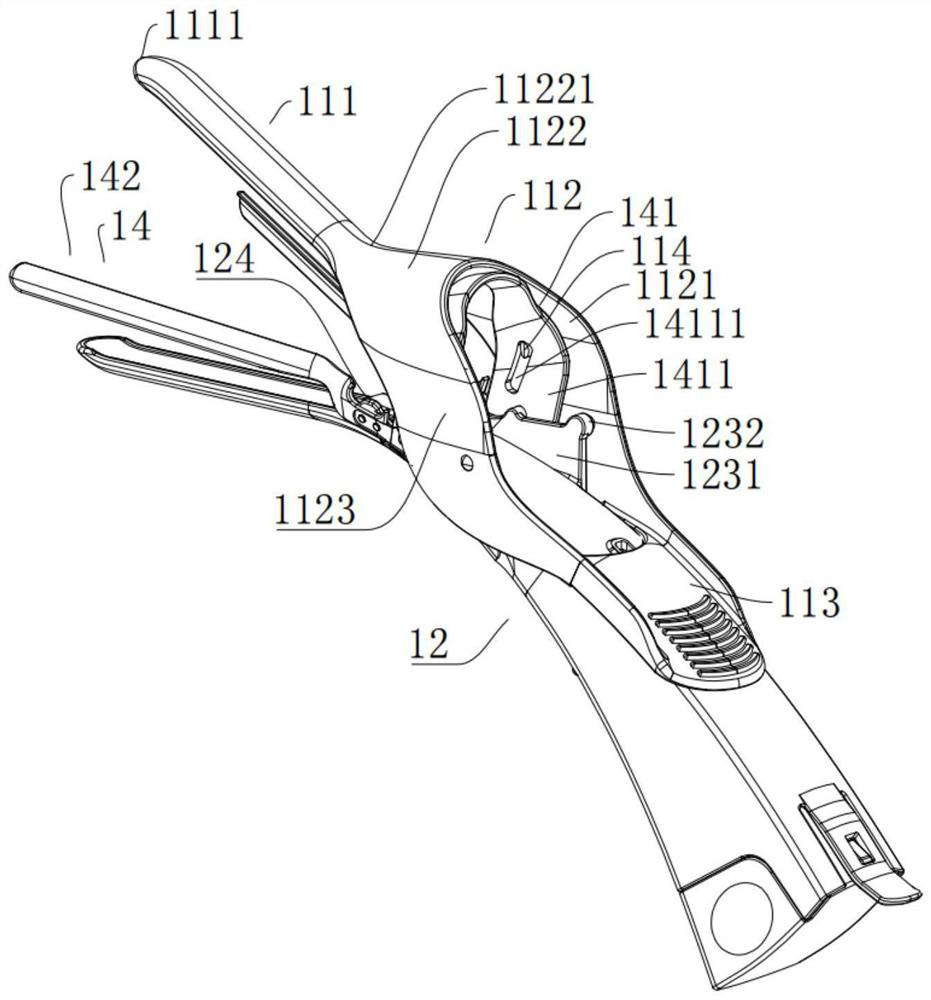
[0075] Continue to refer to figure 1 , figure 2 , image 3 , Figure 4 , Figure 5 as well as Figure 13 , Figure 13 For example Figure 4Enlarged view of part A. The difference between the gynecological examination device in this embodiment and the gynecological examination device in Embodiment 1 is that the expansion mechanism 1 further includes a side expansion assembly 14, and the side expansion assembly 14 includes a side expansion linkage 141 and two side expansion components 142 The side expansion linkage 141 is rotatably connected to the lower expansion assembly 12 and connected to the upper expansion assembly 11. The lower expansion assembly 12 has a side expansion element 124, and one end of the two side expansion elements 142 is connected to the side expansion linkage 141. The other end of each side expansion part 142 extends away from the side expansion linkage part 141 and passes through the side expansion part 124. During the relative opening process of ...

Embodiment 3

[0082] Continue to refer to Figure 16 to Figure 19 , Figure 16 Another cross-sectional view of the inspection mechanism in the embodiment, Figure 17 is another exploded view of the inspection mechanism in the embodiment, Figure 18 For example Figure 16 Enlarged view of Part B, Figure 19 It is a schematic diagram of the structure of the inspection components in the examples. The gynecological examination device in this embodiment is different from the gynecological examination device in Embodiment 1 in that: the see-through part 311 includes a concave lens part 3112, and one end of the concave lens part 3112 is arranged at one end of the first protective part 312, and the concave lens part The other end of 3112 extends toward the inspection component 32 and abuts against the inspection end of the inspection component 32 .

[0083] The setting of the concave lens part 3112 facilitates the maintenance of the state of sticking to the inspection end of the inspection bod...


PUM


Abstract

The invention discloses a gynecologic examination device, which comprises an expansion mechanism, a visual mechanism and an examination mechanism, wherein the expansion mechanism comprises an upper expansion assembly, a lower expansion assembly and a stop assembly, the upper expansion assembly and the lower expansion assembly are connected and can rotate relatively, and the stop assembly comprises a stop piece and a locking piece; the stop piece and the locking piece are arranged on the upper expansion assembly and the lower expansion assembly respectively, the stop piece is detachably connected with the locking piece, the visual mechanism is arranged on the lower expansion assembly, and the examination mechanism is detachably arranged between the upper expansion assembly and the lower expansion assembly. The integrated visual mechanism is used for conducting preliminary visual examination on the vagina, according to the preliminary visual examination result, when a doctor judges that further examination is needed, the examination mechanism directly stretches into the examination channel formed between the upper expansion assembly and the lower expansion assembly, and then further examination and confirmation can be conducted; therefore, one-time diagnosis of gynecological diseases is achieved, the overall examination duration is short, and the screening efficiency is high.

Description

technical field [0001] The invention relates to the technical field of medical devices, in particular to a gynecological examination device. Background technique [0002] Gynecological screening is closely related to the incidence of gynecological diseases. According to the research of Duke University in the United States, women's screening at any time can effectively reduce the lifetime morbidity rate. - 40% reduction, 3 times - 50% reduction. In recent years, the coverage of "two cancers" screening in China has also increased. [0003] In order to improve the screening efficiency of gynecological diseases, medical device products for screening are constantly being innovated and upgraded at home and abroad. The light source and lens are integrated in the vaginal dilator to form a visual module, which can improve the efficiency of preliminary inspection. However, this kind of gynecological medical treatment The improvement of the efficiency of the preliminary inspection of...

Claims


Application Information

Patent Timeline
Patent Type & Authority Applications(China)
IPC IPC(8): A61B1/32A61M31/00A61B1/303A61B1/04
CPCA61B1/32A61M31/00A61B1/303A61B1/04
Inventor 张洋魏信鑫
Owner SUNLAND MEDICAL CO LTD
Why Eureka
Try Eureka