U-Net-based blood vessel image segmentation method, device and equipment

A blood vessel image and blood vessel technology, applied in the field of U-Net-based blood vessel image segmentation, can solve the problems of irrelevant background noise, poor portability, lack of resolution, etc., and achieve the effect of improving segmentation performance

Pending Publication Date: 2021-08-03
GUANGZHOU UNIVERSITY
View PDF7 Cites 1 Cited by
  • Summary
  • Abstract
  • Description
  • Claims
  • Application Information

AI Technical Summary

Problems solved by technology

The main deficiencies of these two types of encoders include: First, there are huge differences in features and imaging principles between medical images and natural images, resulting in poor portability of feature extraction models on these two datasets
[0005] Second, ordinary skip connections in each stage usually directly incorporate local information, which introduces too much irrelevant background noise, making it difficult to distinguish retinal vessels from surrounding mimics and noise, especially tiny vessels
In principle, the high-level features obtained from the deep stage have rich semantic information but lack sufficient resolution, while the low-level features obtained from the shallow stage have rich spatial details but lack global semantic information

Method used


Image

  • U-Net-based blood vessel image segmentation method, device and equipment
  • U-Net-based blood vessel image segmentation method, device and equipment
  • U-Net-based blood vessel image segmentation method, device and equipment

Examples


Embodiment Construction

[0061] In order to make the purpose, technical solution and advantages of the present application clearer, the present application will be further described in detail below in conjunction with the accompanying drawings and embodiments. It should be understood that the specific embodiments described here are only used to explain the present application, and are not intended to limit the present application.

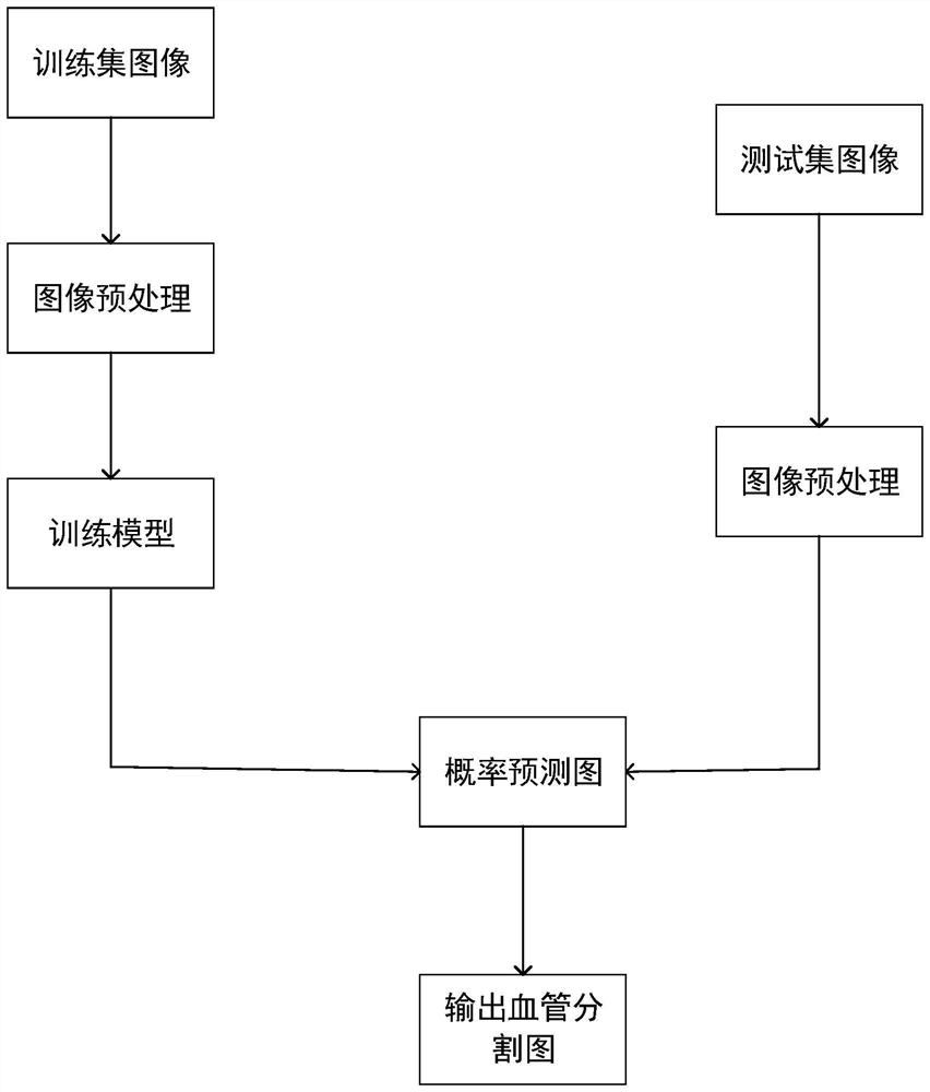
[0062] Aiming at the problems of the prior art, the present invention proposes a U-Net-based blood vessel image segmentation method, including:

[0063] Obtain the blood vessel segmentation dataset;

[0064] Preprocessing the blood vessel segmentation data set;

[0065] Carry out an image block cropping operation on the preprocessed blood vessel segmentation image to obtain sample data;

[0066] According to the sample data, a blood vessel image segmentation network is built through the Pytorch deep learning framework;

[0067] Carrying out blood vessel image segmentati...


PUM


Abstract

The invention discloses a blood vessel image segmentation method, device and equipment based on U-Net. The method comprises the following steps: acquiring a blood vessel segmentation data set; preprocessing the blood vessel segmentation data set; performing image block cutting operation on the preprocessed blood vessel segmentation image to obtain sample data; according to the sample data, building a blood vessel image segmentation network through a Pytorch deep learning framework; and performing blood vessel image segmentation according to the blood vessel image segmentation network, and evaluating a blood vessel image segmentation result. A convolution block in a tube image segmentation network is replaced by a multi-scale feature aggregation block; the first input of the multi-scale feature aggregation block is a multi-scale high-level feature, and the second input of the multi-scale feature aggregation block is a multi-scale low-level feature; according to the blood vessel image segmentation network, the multi-scale high-level features and the multi-scale low-level features in the multi-scale feature aggregation block are fused through the MS-CAM module, the segmentation performance can be improved, and the blood vessel image segmentation network can be widely applied to the technical field of artificial intelligence.

Description

technical field [0001] The invention relates to the technical field of artificial intelligence, in particular to a U-Net-based blood vessel image segmentation method, device and equipment. Background technique [0002] The visible structure of retinal blood vessels can be indicative of many diseases. Accurate segmentation helps capture visible changes in the retinal vascular structure, which helps doctors diagnose eye-related diseases. Therefore, it is particularly important in current retinal image analysis tasks. For example, hypertensive retinopathy is a disease of the retina caused by high blood pressure, and increased curvature or narrowing of blood vessels can be found in patients with high blood pressure. Traditionally, manual segmentation is performed by experts, which is laborious, time-consuming, and suffers from inter-expert subjectivity. In clinical practice, there is a high demand for automatic segmentation methods in order to improve efficiency and reliabili...

Claims


Application Information

Patent Timeline
IPC IPC(8): G06T7/10G06T7/00G06T5/00G06T5/40G06T7/90G06K9/46G06K9/62G06N3/04G06N3/08
CPCG06T7/10G06T7/0012G06T5/40G06T7/90G06N3/08G06T2207/30101G06T2207/20132G06T2207/30041G06T2207/10024G06V10/464G06N3/048G06N3/045G06F18/214G06F18/253G06T5/92
Inventor 彭凌西李动员肖鸿鑫张一梵彭绍湖董志明
Owner GUANGZHOU UNIVERSITY
Why Eureka
Try Eureka