Supercharge Your Innovation With Domain-Expert AI Agents!

Structured light image enhancement method for intraoral 3D reconstruction

An image enhancement and structured light technology, applied in the field of optical 3D scanning, can solve the problems of reducing the number of 3D point clouds, losing 3D detail information, etc., to achieve the effect of enhancing consistency, enhancing 3D details, and increasing the number

Active Publication Date: 2022-07-29
苏州喆安医疗科技有限公司
View PDF7 Cites 0 Cited by
  • Summary
  • Abstract
  • Description
  • Claims
  • Application Information

AI Technical Summary

Problems solved by technology

In turn, the number of reconstructed 3D point clouds is greatly reduced, and the three-dimensional detail information is lost.

Method used


Image

  • Structured light image enhancement method for intraoral 3D reconstruction
  • Structured light image enhancement method for intraoral 3D reconstruction
  • Structured light image enhancement method for intraoral 3D reconstruction

Examples


Embodiment Construction

[0037] The specific embodiments of the present invention will be described below with reference to the accompanying drawings and embodiments.

[0038]

[0039] This embodiment provides a structured light image enhancement method for intraoral 3D reconstruction, which is used to enhance a structured light image obtained by scanning with an oral scanner.

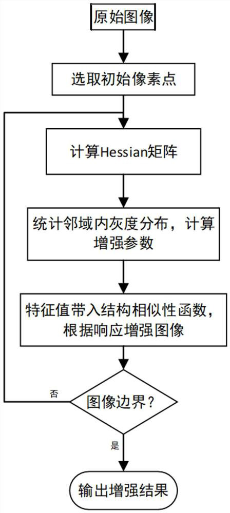
[0040] figure 1 This is a flowchart of the structured light image enhancement method in the embodiment of the present invention.

[0041] Combine the following figure 1 The flow of the structured light image enhancement method for intraoral 3D reconstruction in the embodiment of the present invention will be described.

[0042] Step S1, subtracting the structured light images of adjacent frames to extract fringes.

[0043] Step S2, using the adaptive Hessian image enhancement method to enhance the stripes.

[0044] Wherein, step S2 includes the following sub-steps:

[0045] Step S2-1, select an initial window on the str...


PUM


Abstract

The invention provides a structured light image enhancement method for intraoral 3D reconstruction, which belongs to the technical field of optical three-dimensional scanning and is used for enhancing structured light images scanned by an oral scanner. It is characterized in that it includes the following steps: step S1, subtracting the structured light fringe images of adjacent frames to extract the fringes; step S2, using the adaptive Hessian image enhancement method to enhance the fringes. For the 3D reconstructed fringe structured light image, the similarity function of fringe structure is adjusted dynamically and adaptively to achieve the best fringe enhancement effect. That is to say, based on the anisotropic features extracted by the Hessian matrix, the fringe similarity function is adaptively adjusted by calculating the grayscale statistical features in a small area of ​​the current pixel. To achieve the enhancement effect of the consistency of different brightness and contrast areas. Greatly increases the number of valid 3D point clouds and enhances 3D details.

Description

technical field [0001] The invention belongs to the technical field of optical three-dimensional scanning, and in particular relates to a structured light image enhancement method for intraoral 3D reconstruction. Background technique [0002] With the improvement of social living standards, people's attention to dental health is gradually increasing. When a dentist performs orthodontics or designs dentures on a patient, the first step is to obtain a model of the patient's teeth. The traditional impression method requires the patient to bite the mold on the impression plate first, and then pour the restorative material (such as plaster), and the whole process relies on the manual operation of the dental technician. This method has the disadvantages of long model making time, low precision, and poor patient experience. shortcoming. [0003] In the 1980s, digital impression technology began to be introduced into the field of orthodontics. Commonly used non-contact optical 3D...

Claims


Application Information

Patent Timeline
Patent Type & Authority Patents(China)
IPC IPC(8): G06T5/00
CPCG06T5/005G06T2207/20172G06T2207/10028
Inventor 陈胜杜威
Owner 苏州喆安医疗科技有限公司
Features
  • R&D
  • Intellectual Property
  • Life Sciences
  • Materials
  • Tech Scout
  • Unparalleled Data Quality
  • Higher Quality Content
  • 60% Fewer Hallucinations
Learn More