Non-invasive liver epithelioid vascular smooth muscle lipoma image classification device based on radiomics

A technology of vascular smooth muscle and radiomics, applied in the field of medical image processing

Inactive Publication Date: 2021-08-17
ZHEJIANG UNIV
View PDF1 Cites 8 Cited by
  • Summary
  • Abstract
  • Description
  • Claims
  • Application Information

AI Technical Summary

Problems solved by technology

[0002] Currently, the preoperative differential assessment of liver tumors remains a challenging medical problem for clinicians

Method used


Image

  • Non-invasive liver epithelioid vascular smooth muscle lipoma image classification device based on radiomics
  • Non-invasive liver epithelioid vascular smooth muscle lipoma image classification device based on radiomics
  • Non-invasive liver epithelioid vascular smooth muscle lipoma image classification device based on radiomics

Examples


Embodiment Construction

[0017] The method of the present invention will be further described below in conjunction with the accompanying drawings.

[0018] A radiomics-based non-invasive device for image classification of hepatic epithelioid angiomyolipoma, including:

[0019] Sampling module to obtain CT or MRI image data of patients with confirmed epithelioid angiomyolipoma, liver cancer, or focal nodular hyperplasia of the liver; all epithelioid angiomyolipoma cases are classified as epithelioid angiomyolipoma All cases of liver cancer and focal nodular hyperplasia of the liver were classified into the non-epithelial angiomyolipoma group, and the actual data labels of the cases were given according to the grouping;

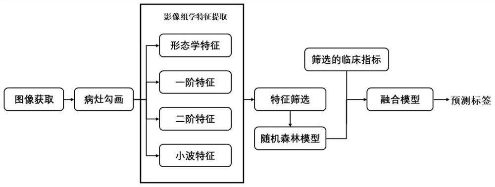
[0020] The lesion area extraction module is used to extract the image of the lesion area of ​​liver epithelioid angiomyolipoma, liver cancer and focal nodular hyperplasia of the liver;

[0021] The feature extraction module is used to extract four types of radiomics features of the im...


PUM


Abstract

The invention discloses a non-invasive liver epithelioid vascular smooth muscle lipoma image classification device based on radiomics, and belongs to the technical field of medical image processing. The non-invasive liver epithelioid vascular smooth muscle lipoma image classification device includes: a sampling module which for collecting CT/MRI images of liver epithelioid vascular smooth muscle lipoma, liver cancer and liver focal nodule hyperplasia meeting requirements; a focus area extraction module which is used for extracting a focus area; a feature extraction module which is used for carrying out radiomics feature extraction on the focus area; a feature screening module which is used for screening radiomics features; a random forest network training module which is used for training a random forest model to obtain a radiomics label; a clinical index fusion module which is used for fusing the radiomics prediction label and the clinical indexes of the patient and training a multivariate logistic regression model; and a classification module which is used for obtaining a final prediction label in combination with a random forest network and a multivariate logistic regression model so as to realize the classification of the liver epithelioid vascular smooth muscle lipoma images. The device is high in recognition precision, high in recognition speed, safe and stable.

Description

technical field [0001] The invention belongs to the technical field of medical image processing, and in particular relates to a radiomics-based non-invasive liver epithelioid angiomyolipoma image classification device. Background technique [0002] Currently, the preoperative differential assessment of liver tumors remains a challenging medical problem for clinicians. On the one hand, because China has a huge population of liver cirrhosis related to hepatitis B virus, a large number of new cases of liver lesions occur every year. On the other hand, with the increasing popularity of health checkups, clinicians can detect various types of liver masses with the help of non-invasive imaging techniques. Typically, clinicians need to evaluate a large number of cases of liver lesions to implement individualized diagnosis, treatment, and follow-up strategies. [0003] Hepatic epithelioid angiomyolipoma (HEAML) is a rare potentially malignant tumor belonging to the PEComas family, ...

Claims


Application Information

Patent Timeline
Patent Type & Authority Applications(China)
IPC IPC(8): G06K9/62G06K9/32G06K9/46G06T7/00G06T7/62G06K9/00
CPCG06T7/0012G06T7/62G06T2207/10081G06T2207/10088G06T2207/30101G06T2207/30056G06T2207/30096G06T2207/20064G06V10/25G06V10/50G06V2201/032G06F2218/06G06F18/213G06F18/24323G06F18/214
Inventor 丁勇邵嘉源夏靖雯田吴炜陆晨燕
Owner ZHEJIANG UNIV
Why Eureka
Try Eureka