Colorectal cancer pathological image prognosis auxiliary prediction method and system

A colorectal cancer and pathological image technology, applied in the field of image processing, can solve the problems of slowing down the doctor's processing speed, missed diagnosis, and increasing the burden on the doctor, and achieve the effect of accurate patient survival time, convenient operation and use, and time saving.

Active Publication Date: 2021-08-27
SOUTH CHINA UNIV OF TECH
View PDF5 Cites 4 Cited by
  • Summary
  • Abstract
  • Description
  • Claims
  • Application Information

AI Technical Summary

Problems solved by technology

[0004] However, the following problems still exist in the prognosis prediction of pathological images formed by medical imaging equipment: (1) After pathologists obtain pathological images of colorectal cancer, they need experienced pathologists to delineate the tumor area and face Exponentially growing medical images, manual processing is slow and inefficient, and missed diagnoses occur from time to time. When there are many patients, the burden on doctors increases. When doctors draw for a long time, the processing speed of doctors will gradually slow down
(2) The prognosis is predicted only based on small image blocks of the tumor area, which lacks sufficient accuracy

Method used


Image

  • Colorectal cancer pathological image prognosis auxiliary prediction method and system
  • Colorectal cancer pathological image prognosis auxiliary prediction method and system
  • Colorectal cancer pathological image prognosis auxiliary prediction method and system

Examples


Embodiment 1

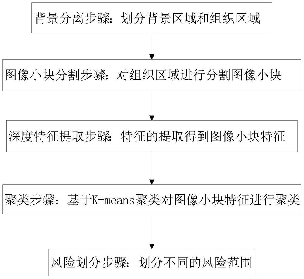
[0055] Such as figure 1 As shown, this embodiment provides a colorectal cancer pathological image prognosis auxiliary prediction method, the method includes the following steps:

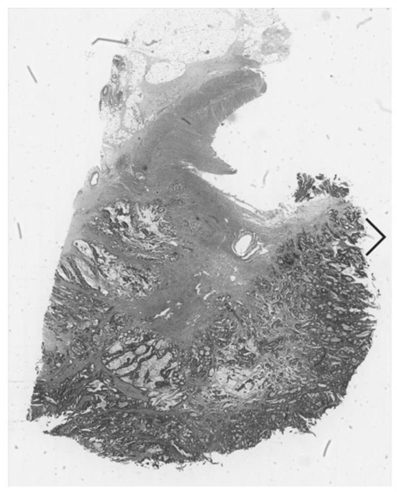
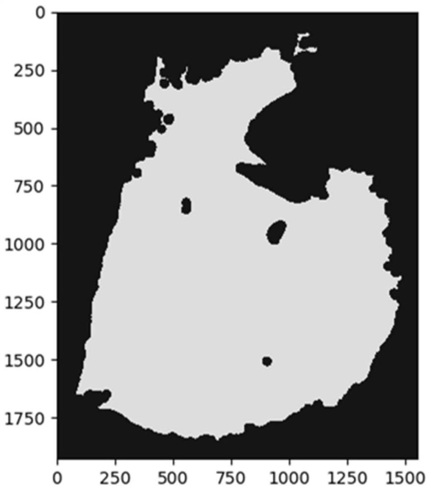
[0056] Background separation steps: eg figure 2 As shown, the pathological image is divided into the tissue area and the background area. First, the tissue area and the background area of ​​the pathological image must be separated to obtain the pathological image. The image format of the pathological image is converted from the RGB space to the HSV color space. The saturation channel part of the image is automatically thresholded and segmented, and then divided into background area and tissue area. In actual application, the default picture format of acquired pathological images is RGB space, where RGB space includes red (R), green (G), blue (B), when the pathological image is converted to HSV (Hue, Saturation, Value) color space , where the HSV color space includes hue (H), saturation (S) and lig...

Embodiment 2

[0074] Through the description of the above embodiments, those skilled in the art can clearly understand that the present invention can be realized by means of software plus a necessary hardware platform, and of course all can be implemented by hardware, but in many cases the former is better implementation. Based on this understanding, all or part of the contribution made by the technical solution of the present invention to the background technology can be embodied in the form of software products, and the computer software products can be stored in storage media, such as ROM / RAM, magnetic disks, optical disks, etc. , including several instructions to make a computer device (which may be a personal computer, a server, or a network device, etc.) execute the method of each embodiment of the present invention or some of the above-mentioned parts of the embodiment.

[0075] This embodiment provides a storage medium, the storage medium may be a storage medium such as ROM, RAM, ma...

Embodiment 3

[0083] Such as Figure 8 As shown, this embodiment provides a colorectal cancer pathological image prognosis auxiliary prediction system, the system includes: a background separation module, an image small block segmentation module, a deep feature extraction module, a clustering module and a risk division module;

[0084] A background separation module for dividing the input pathological image of colorectal cancer into tissue regions and background regions;

[0085] The image small block segmentation module is used to segment the pathological image into small image blocks, and segment the tissue area into small image blocks according to the preset pixel size;

[0086] The deep feature extraction module is used to extract the features of the segmented image blocks according to the convolutional layer and pooling layer of the colorectal cancer survival time prediction model to obtain the image small block features. After the convolutional layer processing, the image small block ...


PUM


Abstract

The invention discloses a colorectal cancer pathological image prognosis auxiliary prediction method and system. The method comprises the following steps: a background separation step: dividing into a background region and a tissue region; an image small block segmentation step: carrying out image small block segmentation on the tissue region according to a preset pixel size; a depth feature extraction step: extracting features of the segmented small image blocks based on a convolutional layer and a pooling layer of a colorectal cancer survival time prediction model to obtain features of the small image blocks, and encoding the small image blocks into a one-dimensional array form after processing of the convolutional layer; a clustering step: clustering the features of the small blocks of the image based on K-means clustering, and dividing tumor epithelial tissues, interstitial tissues, mucus, normal tissues and necrotic parts; and a risk division step: dividing a risk range. According to the invention, the image small blocks of the whole pathological image are automatically classified through K-means clustering, and then different types of image small blocks are selected for joint training, so that the survival time prediction of the prognostic patient of the colorectal cancer patient is more accurate.

Description

technical field [0001] The present invention relates to the technical field of image processing, in particular to an auxiliary prognosis prediction method and system for colorectal cancer pathological images. Background technique [0002] The current prognostic analysis of colorectal cancer mainly requires doctors to carry out pathological TNM staging of colorectal cancer patients, from stage 1 to stage 4 to determine the patient's survival within 5 years. TNM mainly includes tumor infiltration, number of lymph nodes and whether it has metastasized to other organs. The current research on the tumor environment in pathological slices shows that pathology contains a lot of information related to prognosis, and there is a large amount of data waiting to be mined. [0003] A method for predicting the prognosis of pathological images based on deep learning in the prior art requires experienced pathologists to outline the region of interest (region of interest, ROI) in the patholo...

Claims


Application Information

Patent Timeline
Patent Type & Authority Applications(China)
IPC IPC(8): G06T7/00G06T7/10G06K9/62G16H50/20G06N3/08G06N3/04
CPCG06T7/0012G06T7/10G16H50/20G06N3/08G06T2207/30096G06T2207/30028G06N3/045G06F18/23213Y02A90/10
Inventor 覃杰韩楚陈鑫俞祝良刘再毅梁长虹
Owner SOUTH CHINA UNIV OF TECH
Why Eureka
Try Eureka