Breast tumor benign and malignant classification method and device based on ultrasonic image and medium

A breast tumor and ultrasound image technology, applied in the field of medical image processing, can solve the problems of high gray level, inaccurate automatic classification of breast tumor ultrasound images, and difficulty in distinguishing gray-scale similar areas in ultrasound images.

Pending Publication Date: 2021-09-10
HUAQIAO UNIVERSITY
View PDF0 Cites 0 Cited by
  • Summary
  • Abstract
  • Description
  • Claims
  • Application Information

AI Technical Summary

Problems solved by technology

However, the current imaging method of ultrasound images will result in low discrimination between the background and foreground of the image, high and close gray levels, and it is difficult to distinguish the difference between similar gray areas in the ultrasound image. Most studies have not paid

Method used


Image

  • Breast tumor benign and malignant classification method and device based on ultrasonic image and medium
  • Breast tumor benign and malignant classification method and device based on ultrasonic image and medium
  • Breast tumor benign and malignant classification method and device based on ultrasonic image and medium

Examples


Embodiment 1

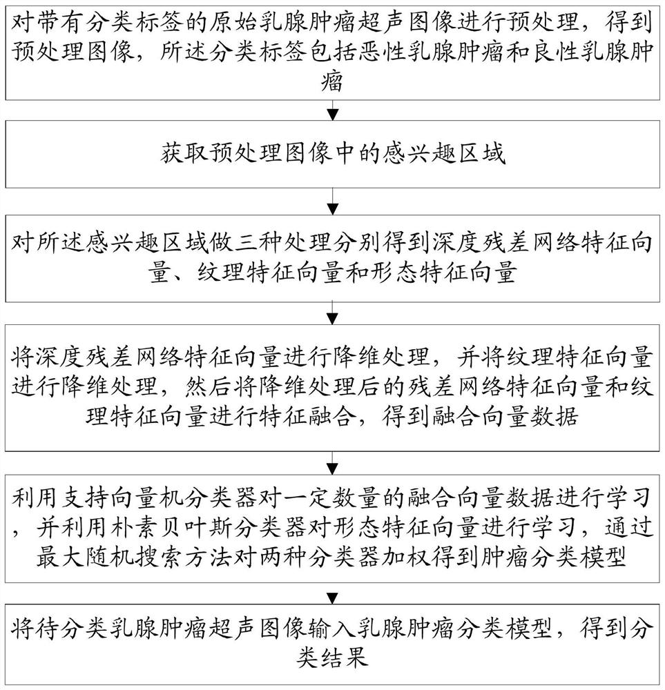
[0079] This embodiment provides a method for classifying benign and malignant breast tumors based on ultrasound images, such as figure 1 As shown, the method includes:

[0080] Step 10, preprocessing the original breast tumor ultrasound image with a classification label to obtain a preprocessed image, and the classification label includes malignant breast tumors and benign breast tumors;

[0081] Step 20, obtaining the region of interest in the preprocessed image;

[0082] Step 30, performing three types of processing on the region of interest to obtain the depth residual network feature vector, texture feature vector and morphological feature vector;

[0083] Step 40, performing dimensionality reduction processing on the deep residual network feature vector, and dimensionality reduction processing on the texture feature vector, and then performing feature fusion on the residual network feature vector and texture feature vector after dimensionality reduction processing, to ob...

Embodiment 2

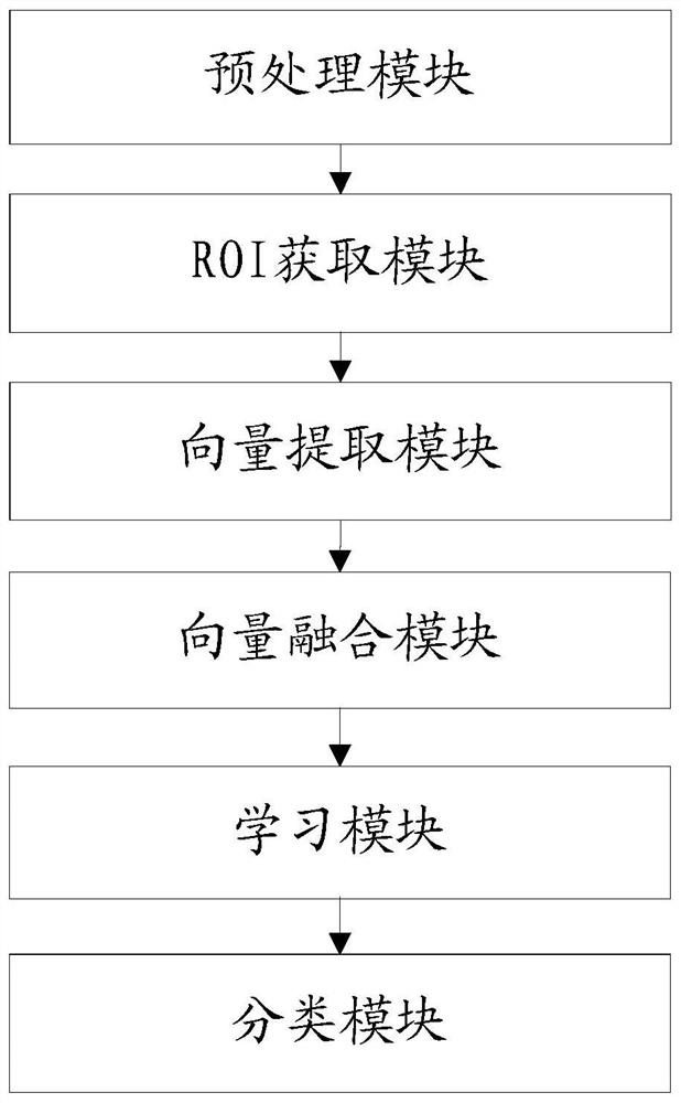
[0130] This embodiment provides a device for classifying benign and malignant breast tumors based on ultrasound images, such as figure 2 As shown, the device includes:

[0131] The preprocessing module is used to preprocess the original breast tumor ultrasound image with a classification label to obtain a preprocessed image, and the classification label includes malignant breast tumors and benign breast tumors;

[0132] The ROI acquisition module is used to acquire the region of interest in the preprocessed image;

[0133] A vector extraction module, configured to perform three types of processing on the region of interest to obtain a depth residual network feature vector, a texture feature vector, and a morphological feature vector;

[0134] The vector fusion module is used to perform dimensionality reduction processing on the deep residual network feature vector, and perform dimensionality reduction processing on the texture feature vector, and then perform feature fusion ...

Embodiment 3

[0180] This embodiment provides a computer-readable storage medium, such as image 3 As shown, a computer program is stored thereon, and when the computer program is executed by a processor, any implementation manner in the first embodiment can be realized.

[0181] Those skilled in the art should understand that the embodiments of the present invention may be provided as methods, apparatuses, or computer program products. Accordingly, the present invention can take the form of an entirely hardware embodiment, an entirely software embodiment, or an embodiment combining software and hardware aspects. Furthermore, the present invention may take the form of a computer program product embodied on one or more computer-usable storage media (including but not limited to disk storage, CD-ROM, optical storage, etc.) having computer-usable program code embodied therein.


PUM


Abstract

The invention provides a breast tumor benign and malignant classification method and device based on an ultrasonic image and a medium, and the method comprises the steps: carrying out the preprocessing of an original breast tumor ultrasonic image with a classification label, and obtaining a preprocessed image; obtaining a region of interest in the preprocessed image; performing three kinds of processing on the region of interest to respectively obtain a deep residual network feature vector, a texture feature vector and a morphological feature vector; carrying out dimension reduction processing on the deep residual network feature vector and the texture feature vector, and then carrying out feature fusion on the deep residual network feature vector and the texture feature vector to obtain fusion vector data; learning a certain amount of fusion vector data by using a support vector machine classifier, learning morphological feature vectors by using a naive Bayes classifier, and obtaining a tumor classification model by weighting the two classifiers; and inputting the breast tumor ultrasonic image to be classified into the breast tumor classification model to obtain a classification result. By adopting the method provided by the invention, automatic classification of the breast tumor ultrasonic images can be realized more accurately.

Description

technical field [0001] The invention relates to the field of medical image processing, in particular to a method, device and medium for benign and malignant breast tumors based on ultrasound images. Background technique [0002] Cancer is one of the most important diseases threatening human health worldwide. According to the statistics of the World Health Organization, the incidence of cancer has been on the rise. Early detection, early diagnosis and early treatment are the key to improving the cure rate of cancer and reducing the mortality rate. Ultrasound imaging technology has been widely used in the detection of breast tumors due to its versatility, safety and high sensitivity. Thyroid Ultrasound, Breast Ultrasound, Prostate Ultrasound, Cardiac Ultrasound and Renal Ultrasound, etc. [0003] At present, ultrasound is the preferred imaging examination and preoperative evaluation method for the diagnosis of breast tumors. However, correct interpretation of breast ultrasou...

Claims


Application Information

Patent Timeline
IPC IPC(8): G16H50/20G06K9/46G06K9/62
CPCG16H50/20G06F18/2411G06F18/24155
Inventor 杜永兆刘卫郭明辉柳培忠
Owner HUAQIAO UNIVERSITY
Why Eureka
Try Eureka