
X-ray section imaging method, storage medium and imaging system

An imaging method and imaging system technology, applied in the field of X-rays, can solve the problems of limited flexibility and application scenarios, difficulty and high manufacturing cost of X-ray slice imaging systems, and restrictions on popularization and application of X-ray slice imaging systems , to achieve the effects of easy manufacturing and mass deployment, obvious price competitive advantage, timeliness and universal applicability

Pending Publication Date: 2021-10-22
BEIJING YOUTONG SHANGHAO TECH CO LTD
View PDF0 Cites 0 Cited by
  • Summary
  • Abstract
  • Description
  • Claims
  • Application Information

AI Technical Summary

Problems solved by technology

[0005] In order to ensure that the main frame of the X-ray imaging system, the high-precision guide rail that slides with the main frame, and the fixed track for the movement of the X-ray source meet high geometric accuracy requirements, the above-mentioned mechanical equipment needs to be made of high-cost materials, such as high-strength aviation aluminum At the same time, high-precision processing equipment and strict processing technology are required, which makes the manufacturing difficulty and manufacturing cost of the existing X-ray section imaging system remain high, which greatly limits the popularization and application of X-ray section imaging system products.
At the same time, the high-precision requirements of the mechanical structure also limit the flexibility and application scenarios of the mechanical structure design of the equipment.
[0006] In addition, for many products that have been used in hospitals, the accuracy of the main frame of the X-ray imaging system, the guide rails that match it, and the fixed orbit of the X-ray source movement are low. Imaging system, in the imaging process, mechanical errors directly affect the accuracy of the shooting position of the X-ray source, which in turn affects the imaging quality
If the relevant mechanical structure is upgraded with high precision, high transformation costs will be incurred, the meaning of transformation will be lost, and the need for upgrading and transformation of existing X-ray products in underdeveloped areas will not be met.

Method used


Image

  • X-ray section imaging method, storage medium and imaging system
  • X-ray section imaging method, storage medium and imaging system
  • X-ray section imaging method, storage medium and imaging system

Examples


Embodiment Construction

[0054] In order to explain the overall concept of the present application more clearly, the following detailed description will be given by way of examples in combination with the accompanying drawings.

[0055] In the following description, many specific details are set forth in order to fully understand the application, but the application can also be implemented in other ways different from those described here, therefore, the protection scope of the application is not limited by the specific details disclosed below. EXAMPLE LIMITATIONS.

[0056] In addition, the terms "first" and "second" are used for descriptive purposes only, and cannot be interpreted as indicating or implying relative importance or implicitly specifying the quantity of indicated technical features. Thus, a feature defined as "first" and "second" may explicitly or implicitly include one or more of these features.

[0057] X-ray imaging technology includes digital radiography (DR-Digital Radiography). Th...


PUM


Abstract

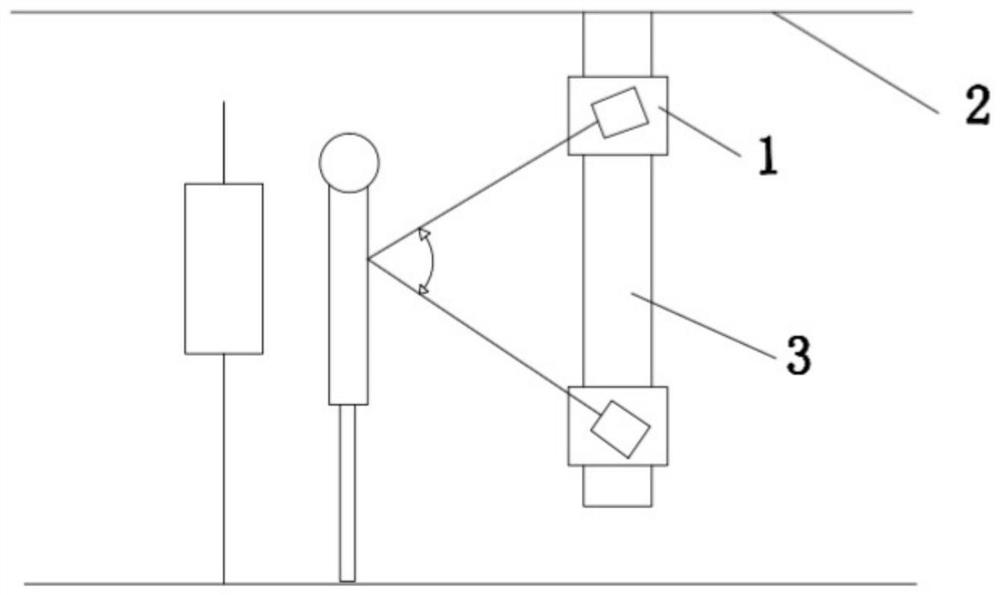
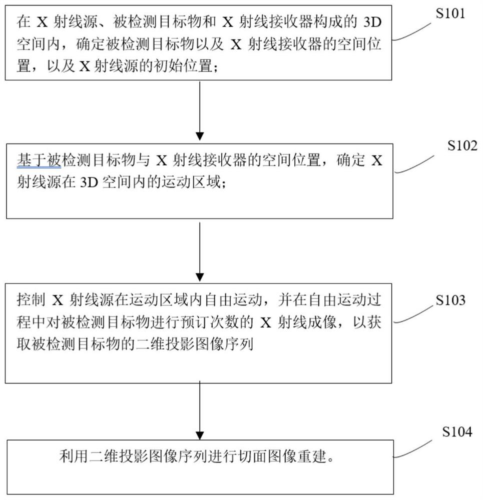
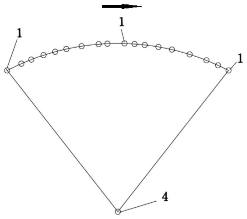
The invention provides an X-ray section imaging method which comprises the following steps: determining the spatial positions of a detected target object and an X-ray receiver and the initial position of an X-ray source in a 3D space formed by the X-ray source, the detected target object and the X-ray receiver; determining a motion area of the X-ray source in the 3D space based on the spatial positions of the detected target object and the X-ray receiver; controlling the X-ray source to move freely in the movement area, performing X-ray imaging on the detected target object for a preset number of times in the free movement so as to obtain a two-dimensional projection image sequence of the detected target object, and utilizing the two-dimensional projection image sequence to reconstruct a section image sequence; and the invention also provides a a storage medium and an imaging system. The method has the beneficial effects that the X-ray imaging system freely moves in a selected space motion area, X-ray image data of a detected target object can be acquired at any projection angle or any SID, and the section image can be reconstructed without being limited by the precision of mechanical parts in the X-ray imaging system and the space motion trail.

Description

technical field [0001] The invention belongs to the technical field of X-rays, and in particular relates to an X-ray section imaging method, a storage medium and an imaging system. Background technique [0002] X-ray imaging technology is an image diagnosis method with the largest usage, the widest application area and the largest number of equipment among various medical imaging technologies, and has extremely high clinical value. It is widely used in imaging diagnosis and image-guided therapy in medical and veterinary fields , and is also widely used in the field of industrial non-destructive testing. [0003] In traditional X-ray imaging systems, X-rays exhibit different attenuation characteristics when passing through materials with different densities. The density difference information of the internal structure of the imaged object is obtained by detecting the attenuation degree of X-rays, thereby generating X-ray images. In such conventional X-ray imaging systems, an...

Claims


Application Information

Patent Timeline
Patent Type & Authority Applications(China)
IPC IPC(8): A61B6/00
CPCA61B6/4411A61B6/4476A61B6/5211A61B6/582
Inventor 邹鲁民
Owner BEIJING YOUTONG SHANGHAO TECH CO LTD
Learn More