Radiotherapy dose prediction method and device
A dose prediction and dose technology, applied in radiation therapy, X-ray/γ-ray/particle irradiation therapy, treatment, etc., can solve the problems of difficult dose calculation and optimization, long time, etc.
- Summary
- Abstract
- Description
- Claims
- Application Information
AI Technical Summary
Problems solved by technology
Method used
Image

Examples
no. 1 example 1
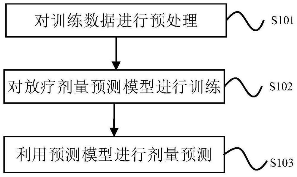
[0023] figure 1 It is a schematic flowchart of a radiotherapy dose prediction method according to the first embodiment of the present invention, as shown in figure 1 As shown, the method includes the following three steps.
[0024] Step S101: Preprocessing the training data. According to the patient's medical image, the patient phantom is established; the dose distribution H of radioactive particles in the patient phantom is calculated using the first calculation method of radiotherapy dose; the dose distribution L1 in the uniform water phantom is calculated using the TG-43 method, and the tissue difference is used The qualitative correction method corrects the dose distribution L1 to obtain the dose distribution L of radioactive particles in the patient phantom; the position of the radioactive particles is modified several times, and the dose distributions H and L are recalculated each time the position is modified.
[0025] Exemplarily, the patient phantom may be three-dim...
no. 1 example
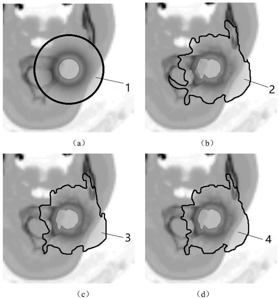
[0032] figure 2 It is a schematic diagram of dose distribution of a radiotherapy dose prediction method according to the first embodiment of the present invention. Such as figure 2 as shown, figure 2 (a) is a schematic diagram of the dose distribution of radioactive particles in the water model calculated by the TG-43 method; figure 2 (b) is figure 2 (a) Schematic diagram of dose distribution after dose distribution correction. Since the TG-43 method directly obtains the radioactive particles in the uniform water model, therefore figure 2 The dose distribution 1 in (a) is a uniform circle, and the dose distribution after heterogeneity correction is as follows figure 2 The dose distribution in (b) is shown in 2.
[0033] In an optional embodiment, the first calculation method of the radiotherapy dose is a Monte Carlo simulation method. Monte Carlo simulation calculates the dose distribution based on the computer simulation of the physical process of the particles ...
Embodiment 2
[0041] The embodiment of the present invention provides a radiotherapy dose prediction device, which is mainly used to implement the radiotherapy dose prediction method provided in the above-mentioned content of the embodiment of the present invention. The radiotherapy dose prediction device provided by the embodiment of the present invention will be described in detail below.
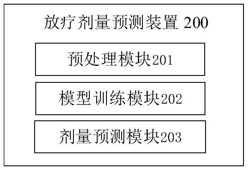
[0042] image 3 It is a structural schematic diagram of a radiotherapy dose prediction device according to the second embodiment of the present invention. Such as image 3 As shown, the radiotherapy dose prediction device 200 includes the following modules:
[0043] Preprocessing module 201, which is used to establish a patient phantom according to the patient's medical image; calculate the dose distribution H of radioactive particles in the patient phantom by using the first calculation method of radiotherapy dose; use the TG-43 method to calculate the dose distribution H in the uniform water phantom...
PUM
 Abstract
Description
Claims
Application Information
PatSnap Eureka turns technology decisions into work you can execute. Powered by our Innovation Knowledge Graph, it runs expert workflows across engineering, life sciences, materials and intellectual property. Get your review-ready output in minutes.


