Eye fundus image analysis method and system and electronic equipment

A fundus image and analysis method technology, applied in the field of medical data analysis, can solve problems such as difficult to meet the needs of disease course monitoring, difficult to accurately quantify leopard-shaped fundus, and high cost

Pending Publication Date: 2021-12-10
BEIJING AIRDOC TECH CO LTD +1
View PDF16 Cites 6 Cited by
  • Summary
  • Abstract
  • Description
  • Claims
  • Application Information

AI Technical Summary

Problems solved by technology

[0008] When the ophthalmologist directly observes the fundus image, the difference between the leopard pattern and other areas of the fundus is small when the severity of the leopard pattern is low. To what extent is it serious, it will take a lot of time and cost
Moreover, the presence of leopard prints in different areas of the fundus and different degrees of leopard prints have different effects on vision. It is difficult for ophthalmologists to quickly and accurately give the screening results with the naked eye.
These problems are mainly due to the existence of leopard-shaped fundus that is difficult to accurately quantify, so it is difficult to meet the needs of continuous disease course monitoring

Method used


Image

  • Eye fundus image analysis method and system and electronic equipment
  • Eye fundus image analysis method and system and electronic equipment
  • Eye fundus image analysis method and system and electronic equipment

Examples


Embodiment Construction

[0032] In order to make the object, technical solution and advantages of the present invention clearer, the present invention will be further described in detail below through specific embodiments in conjunction with the accompanying drawings. It should be understood that the specific embodiments described here are only used to explain the present invention, not to limit the present invention.

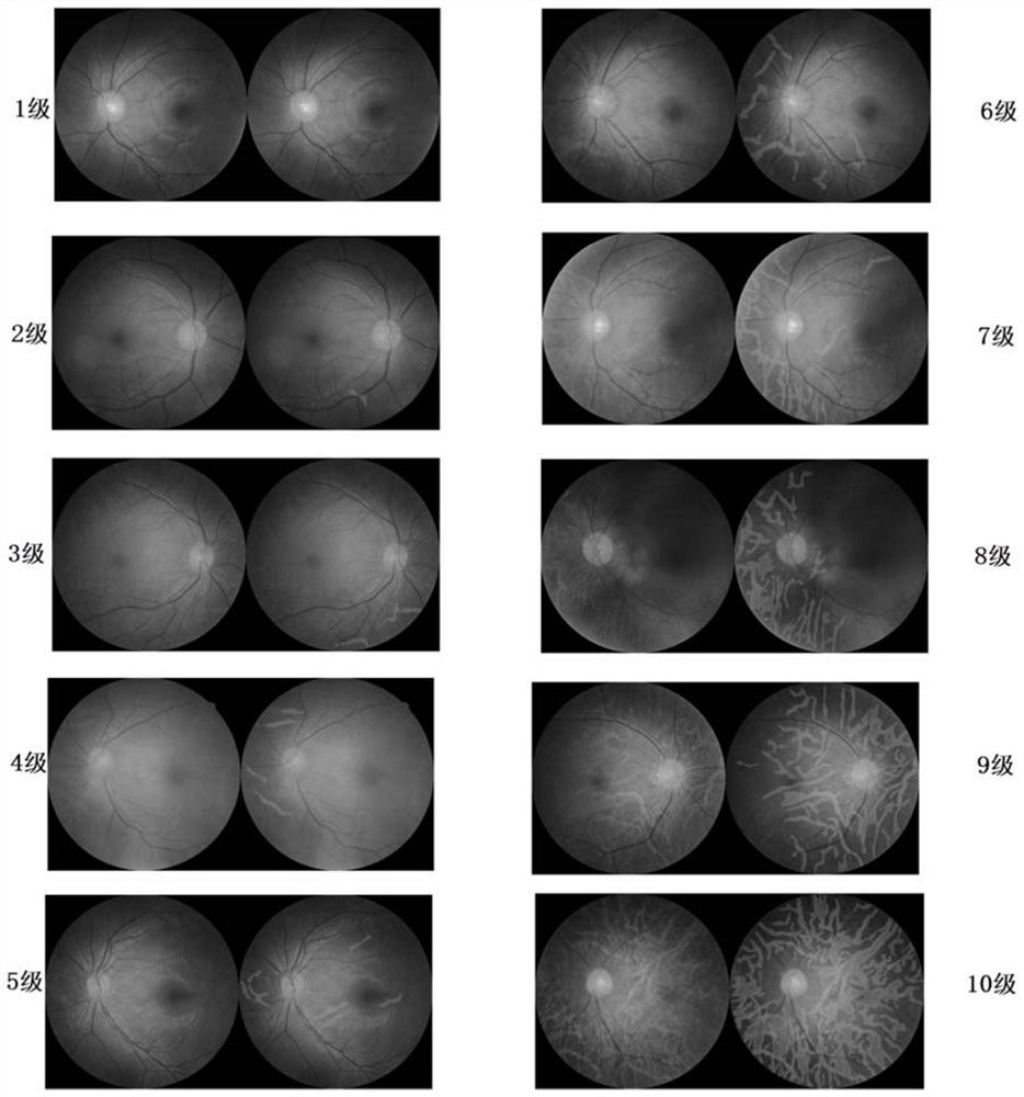
[0033] As mentioned in the background technology section, when the ophthalmologist directly observes the fundus image, the difference between the leopard print and other areas of the fundus is small when the severity of the leopard print is low, and it is difficult for the naked eye to quickly distinguish the leopard print from other areas of the fundus. When the degree is high, it is difficult to quickly determine the specific degree of seriousness, and it will take a lot of time and cost. Moreover, the appearance of leopard prints in different areas of the fundus and different degree...


PUM


Abstract

The embodiment of the invention provides an eye fundus image analysis method and system and electronic equipment. The eye fundus image analysis method comprises the following steps: acquiring a to-be-analyzed eye fundus image; performing pixel-level segmentation on the eye fundus image to obtain leopard print segmentation information which identifies that each pixel belongs to a leopard print region or a non-leopard print region; based on the eye fundus image, segmenting a plurality of regions of interest with different degrees of influence of the leopard print on vision, and obtaining region segmentation information for identifying the region of interest to which each pixel belongs; and performing quantitative analysis on the eye fundus leopard print according to the leopard print segmentation information and the region segmentation information. According to the method, quantitative analysis is carried out on the eye fundus leopard print more accurately according to the leopard print segmentation information and the region segmentation information, so that a diagnosis result can be given quickly and accurately; and a doctor does not need to spend a lot of energy and time to learn and analyze how to determine the degree of the leopard print according to the eye fundus image.

Description

technical field [0001] The present invention relates to the field of medical data analysis, in particular to analyzing fundus images by using a neural network model, and more specifically, to a fundus image analysis method, system and electronic equipment. Background technique [0002] The fundus is the area at the inner back of the eye that includes anatomical structures such as the retina, optic disc, macula, and central retinal artery. Usually, the fundus camera can be used to observe the condition of the fundus non-invasively, painlessly and quickly, and make a diagnosis of related diseases. Because of its convenience and non-destructive characteristics, and because various diseases of the human body can be reflected on the fundus, disease screening through fundus images has become an important means for the diagnosis of diabetes, glaucoma and other diseases and the management of chronic diseases. [0003] Leopard-like fundus refers to the appearance of leopard-like tex...

Claims


Application Information

Patent Timeline
Patent Type & Authority Applications(China)
IPC IPC(8): A61B3/12A61B3/14G06K9/62G06N3/04G06N3/08G06T7/00G06T7/11
CPCA61B3/12A61B3/14A61B3/0025G06T7/0012G06T7/11G06N3/04G06N3/08G06T2207/30041G06T2207/20081G06F18/241
Inventor 王璘杨志文贺婉佶王欣琚烈戈宗元王斌赵昕和超陈羽中张大磊
Owner BEIJING AIRDOC TECH CO LTD
Why Eureka
Try Eureka