Computer-assisted breast cancer pathological image diagnosis method based on artificial intelligence

A computer-aided, pathological image technology, applied in computer-aided medical procedures, computer parts, calculations, etc., can solve problems such as subjectivity and low efficiency, and achieve the effect of avoiding subjective judgment, improving work efficiency, and reducing workload.

Pending Publication Date: 2022-03-11
PEKING UNION MEDICAL COLLEGE HOSPITAL CHINESE ACAD OF MEDICAL SCI +1
View PDF0 Cites 1 Cited by
  • Summary
  • Abstract
  • Description
  • Claims
  • Application Information

AI Technical Summary

Problems solved by technology

[0006] The purpose of the present invention is to solve the problems of subjectivity and low efficiency in the existing manual interpretation of pathological subtypes of breast cancer

Method used


Image

  • Computer-assisted breast cancer pathological image diagnosis method based on artificial intelligence
  • Computer-assisted breast cancer pathological image diagnosis method based on artificial intelligence
  • Computer-assisted breast cancer pathological image diagnosis method based on artificial intelligence

Examples


Embodiment 1

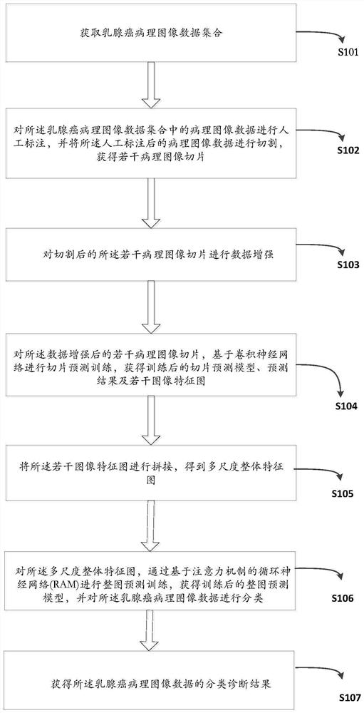
[0026] Embodiment 1: Carry out model training on the pathological image set of breast cancer, and obtain the trained slice prediction model and whole image prediction model. Such as figure 1 Shown, the method for model training of the present invention is as follows:

[0027] Step 1: Acquire breast cancer pathological image data set. The pathological image set includes images of different sizes of the original image of the same breast cancer pathological slice.

[0028]In this embodiment, pathological full-field slice images of breast cancer are used. The dataset includes invasive lobular carcinoma (ILC), invasive micropapillary carcinoma (IMPC), tubular carcinoma (TC / ICC), mucinous carcinoma (MC), apocrine gland carcinoma (AC) and invasive ductal carcinoma (IDC) Six breast cancer subtypes, with different breast cancer subtypes defined by distinct morphological patterns.

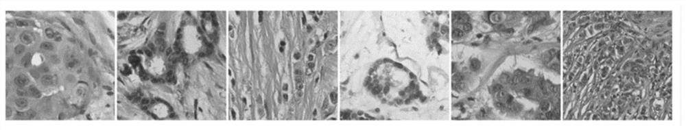
[0029] Such as image 3 As shown, the data set was produced by digitizing breast cancer histopatholo...

Embodiment 2

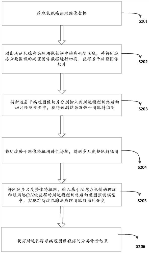
[0050] Embodiment 2: Based on the trained slice prediction model and whole image prediction model, breast cancer diagnosis and classification are performed on breast cancer pathological images. Such as figure 2 Shown, the method for image diagnosis of the present invention is as follows:

[0051] Step 1: Obtain pathological image data of breast cancer. In this embodiment, pathological full-field slice images of breast cancer are used. The dataset includes invasive lobular carcinoma (ILC), invasive micropapillary carcinoma (IMPC), tubular carcinoma (TC / ICC), mucinous carcinoma (MC), apocrine gland carcinoma (AC) and invasive ductal carcinoma (IDC) Six breast cancer subtypes, with different breast cancer subtypes defined by distinct morphological patterns.

[0052] Such as image 3 As shown, the data set was produced by digitizing breast cancer histopathological slides with a slide scanner with a maximum magnification of 40× (0.2 μm / pixel) using hematoxylin-eosin staining (...


PUM


Abstract

The invention provides a computer-aided breast cancer pathology image diagnosis method based on artificial intelligence, and the method comprises the steps: carrying out the model training of a breast cancer pathology image set, and obtaining a trained slice prediction model and a whole image prediction model; and secondly, based on the trained slice prediction model and the whole image prediction model, performing breast cancer diagnosis classification on the breast cancer pathological image. According to the method, the trained neural network can be used for layering a large number of breast cancer pathology images at different positions, the data processing speed and accuracy are greatly improved, and the problems of subjectivity, low efficiency and the like existing in existing breast cancer pathology subtype manual interpretation are solved.

Description

technical field [0001] The invention relates to a pathological image diagnosis method, in particular to an artificial intelligence-based computer-assisted breast cancer pathological image diagnosis method, which belongs to the technical field of medical computer image processing. Background technique [0002] Breast cancer is one of the most common malignant tumors in women all over the world, which greatly threatens women's health. The diagnosis of breast cancer mainly depends on pathological diagnosis. [0003] Pathological diagnosis is carried out by observing a microscopic glass specimen called a columnar body through a microscope. The microscope specimen is made of organs, tissues and cells collected from the patient, and the specimen is cut into thin slices several microns thick , Microscopic observation of the stained specimen will become the basis for the doctor to diagnose the disease, and the chemical tissue that will play a role in the judgment of treatment metho...

Claims


Application Information

Patent Timeline
Patent Type & Authority Applications(China)
IPC IPC(8): G16H50/20G16H70/60G06T7/00G06N3/08G06N3/04G06K9/62G06V10/25G06V10/764G06V10/82
CPCG16H50/20G16H70/60G06T7/0012G06N3/08G06T2207/20081G06T2207/30068G06T2207/20104G06N3/045G06F18/2415G06F18/241
Inventor 梁智勇邹昊吴焕文郭玉成李俊杰
Owner PEKING UNION MEDICAL COLLEGE HOSPITAL CHINESE ACAD OF MEDICAL SCI
Why Eureka
Try Eureka