
New coronal pneumonia CT image segmentation method and terminal device

A CT image, pneumonia technology, applied in the field of medical image segmentation, can solve the problem of difficult and accurate segmentation of new crown pneumonia lesions, and achieve the effect of speeding up reasoning

Inactive Publication Date: 2022-03-22
HUNAN NORMAL UNIVERSITY
View PDF13 Cites 0 Cited by
  • Summary
  • Abstract
  • Description
  • Claims
  • Application Information

AI Technical Summary

Problems solved by technology

[0006] The present invention provides a new coronary pneumonia CT image segmentation method and terminal equipment, thereby solving the technical problem that it is difficult to accurately segment the new coronary pneumonia lesion area in the prior art

Method used


Image

  • New coronal pneumonia CT image segmentation method and terminal device
  • New coronal pneumonia CT image segmentation method and terminal device
  • New coronal pneumonia CT image segmentation method and terminal device

Examples


Embodiment Construction

[0061] The technical solutions of the present invention will be further described in detail below in conjunction with the accompanying drawings and specific implementation cases. It should be understood that the following examples are only for illustrating and explaining the present invention, and should not be construed as limiting the protection scope of the present invention. All technologies realized based on the above contents of the present invention are covered within the scope of protection intended by the present invention.

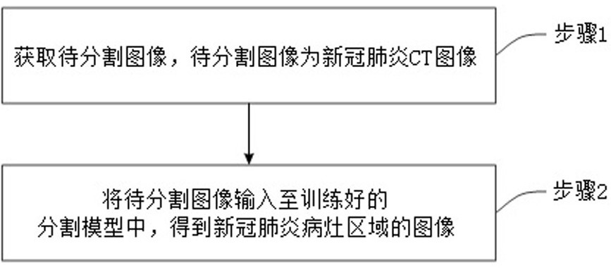
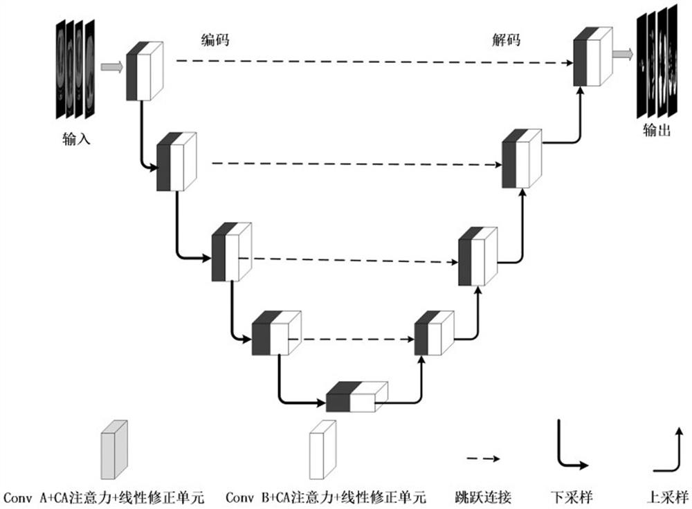
[0062] Such as Figure 1 to Figure 3 As shown, a new coronary pneumonia CT image segmentation method of the present invention is characterized in that it includes:

[0063] Step 1. Obtain the image to be segmented, wherein the image to be segmented is a CT image of COVID-19.

[0064] Step 2. Input the image to be segmented into the trained segmentation model to obtain the image of the lesion area of ​​the new coronary pneumonia.

[0065] In th...


PUM


Abstract

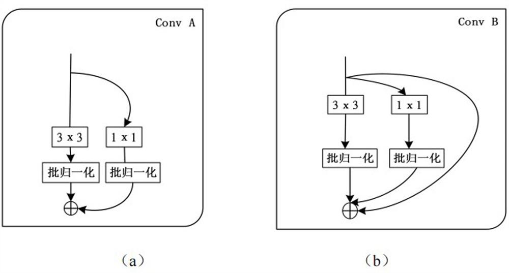
The invention discloses a new coronal pneumonia CT image segmentation method and terminal equipment. The method comprises the following steps: acquiring a to-be-segmented new coronal pneumonia CT image; inputting the image into a trained segmentation model to obtain an image of a new coronal pneumonia focus area; wherein the segmentation model comprises a plurality of down-sampling modules connected in sequence and up-sampling modules corresponding to the down-sampling modules; each sampling module comprises a first extraction unit and a second extraction unit which are connected in sequence; the convolution modules of the two extraction units are structure re-parameterization convolution modules. According to the structural re-parameterization convolution module, a multi-branch structure is used during training, the expression ability of the model is enhanced, a single-path structure is used during reasoning, the reasoning speed is increased, and a diagnosis result is rapidly obtained. Meanwhile, in order to learn hierarchical representation from feature maps with different scales, the model is enhanced to extract image edge information and enable gradient to flow back more quickly, output on each side of up-sampling is connected with a mixed loss function, and pixel-level segmentation of the image is achieved.

Description

technical field [0001] The invention belongs to the technical field of medical image segmentation, and specifically relates to a novel coronavirus pneumonia CT image segmentation method and terminal equipment, especially a novel coronavirus pneumonia CT image segmentation method and terminal equipment based on structural reparameterization and multi-scale dynamic weight loss. Background technique [0002] The new coronavirus is mainly transmitted through the respiratory tract, and can also be transmitted through environmental contact. Studies have shown that COVID-19 is far more than a respiratory disease, but a multi-system disease that can cause damage to multiple human organs including the heart, kidneys, and nervous system. [0003] RT-PCR has been established as the gold standard for COVID-19 screening. RT-PCR (ReverseTranscription-Polymerase Chain Reaction) can obtain viral RNA in specimens through nasopharyngeal swabs, oropharyngeal swabs, bronchoalveolar lavage flui...

Claims


Application Information

Patent Timeline
Patent Type & Authority Applications(China)
IPC IPC(8): G06T7/12G06T7/13G06T7/194G06N3/04G06N3/08
CPCG06T7/12G06T7/13G06T7/194G06N3/08G06T2207/10081G06T2207/20081G06T2207/20084G06T2207/30061G06N3/048G06N3/045
Inventor 刘金平吴娟娟赵刚劲李梦可郑坤一杜保强王靖超陈文祥
Owner HUNAN NORMAL UNIVERSITY
Learn More