Urinography CT image kidney segmentation method and system

A CT imaging and urography technology, applied in the field of medical imaging, can solve the problems of incomplete target domain segmentation results, loss of local information in AdaptSegNet, weakened model segmentation performance, etc., to overcome the problem of gradient disappearance, low computational cost, and high accuracy. sexual effect

Pending Publication Date: 2022-04-22
罗雄彪 +1
View PDF0 Cites 0 Cited by
  • Summary
  • Abstract
  • Description
  • Claims
  • Application Information

AI Technical Summary

Problems solved by technology

However, when the distribution of the test data (target domain) is different from the training data (source domain), the segmentation performance of the model will be weakened, and such problems are generally called do

Method used


Image

  • Urinography CT image kidney segmentation method and system
  • Urinography CT image kidney segmentation method and system
  • Urinography CT image kidney segmentation method and system

Examples


Example Embodiment

[0072] In order to make the objects, technical solutions, and advantages of the present invention more clearly, the technical solutions in the embodiments in the embodiments will be described in contemplation in the embodiment of the present invention, and will be described in connext of the embodiment of the present invention. It is an embodiment of the invention, not all of the embodiments. Based on the embodiments of the present invention, all other embodiments obtained by those of ordinary skill in the art are in the range of the present invention without making creative labor premise.

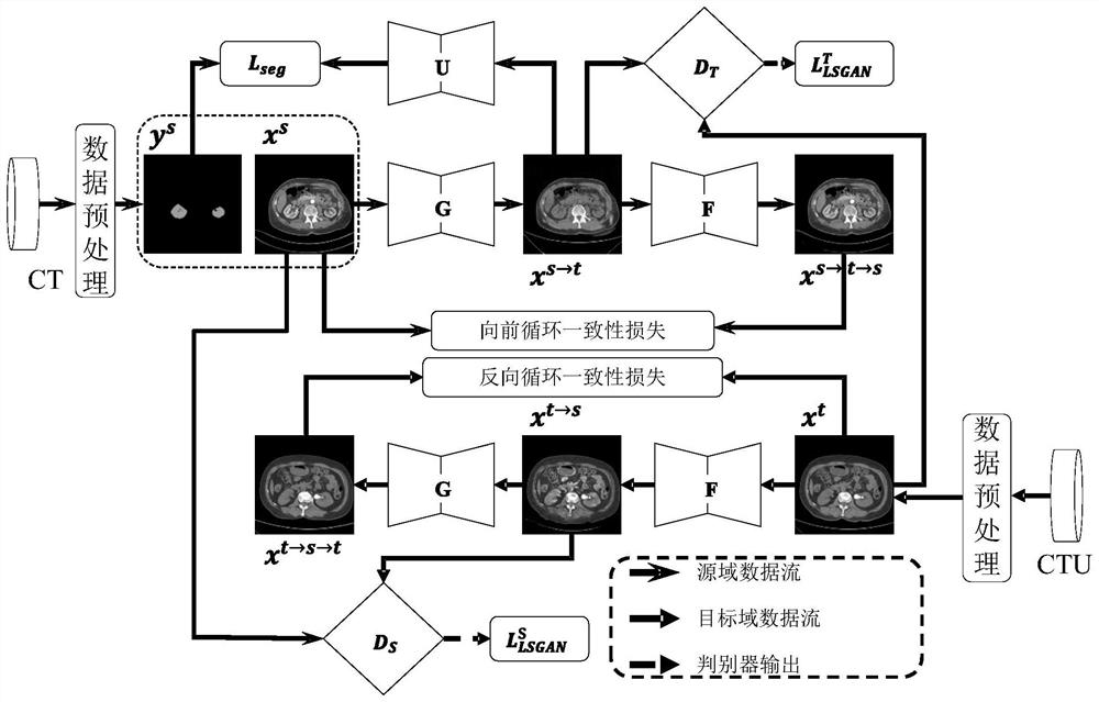
[0073] Examples of the present invention provide a urinary tract angiography CT image kidney segmentation method. Based on the translation and confrontation learning of no supervisory domain, using CT data labeled with kidney, CTU data -free CTU data, and cycle generating network generation to generate the kidney with the kidneys with the kidneys with the kidneys. The labeled CTU data, that is...


PUM


Abstract

The invention provides a urinary tract radiography CT image kidney segmentation method and system. The method comprises the steps that CT data with kidney labels and CTU data without kidney labels are acquired; determining a cyclic generative adversarial network, and generating synthetic CTU data by using the CT data with the kidney label, the CTU data without the kidney label and the cyclic generative adversarial network; determining a segmentation network, and training the segmentation network based on the synthesized CTU data; and marking and segmenting the urinary tract angiography CT image by using the segmentation network. According to the method, the CTU data is synthesized through the cyclic generative adversarial network, the synthesized CTU data based on the CTU data without kidney annotation is automatically obtained, the segmentation network is trained based on the synthesized CTU data, the network credibility is high, the pertinence is high, and the annotation segmentation result of the urinary tract angiography CT image is automatically obtained based on the segmentation network and also has very high accuracy.

Description

technical field [0001] The invention belongs to the technical field of medical imaging, in particular to a kidney segmentation method and system for urography CT images. Background technique [0002] Flexible ureteroscopic lithotripsy is an effective minimally invasive procedure for the treatment of kidney stones. The anatomical structure of the kidney, renal pelvis, and calyces will greatly affect the stone-clearing rate of surgery. Accurate measurement of anatomical parameters, such as the angle between the renal pelvis and the inferior calices, and the diameter and width of the inferior renal calyces, is very important for reducing the risk of flexible ureteroscopic lithotripsy. However, accurate parameter measurement requires three-dimensional segmentation and reconstruction of the kidney based on computed tomography urography (CTU). With the increase of a large amount of medical imaging data and the emergence of high-performance computing equipment, researchers have p...

Claims


Application Information

Patent Timeline
IPC IPC(8): G06T7/10G06N3/04G06N3/08
CPCG06T7/10G06N3/084G06T2207/10081G06T2207/20081G06T2207/20084G06T2207/30084G06N3/045
Inventor 罗雄彪万英
Owner 罗雄彪
Why Eureka
Try Eureka