
Oral cavity supporting equipment for gastroscopy

A supporting device and gastroscopic inspection technology, which is applied in the direction of gastroscope, oral mirror, application, etc., can solve the problems of patients' tooth pain, biting the mouthpiece, and uniform specifications, so as to avoid tooth pain, increase comfort, avoid The effect of resistance

Inactive Publication Date: 2022-04-29
郑州大学第五附属医院
View PDF0 Cites 1 Cited by
  • Summary
  • Abstract
  • Description
  • Claims
  • Application Information

AI Technical Summary

Problems solved by technology

However, the existing oral support appliance is a simple mouthpiece, which has the following problems: 1. The specifications are uniform and cannot be adjusted
During the examination, patients are prone to discomfort, and then they will perform occlusal movements. When the occlusal force is strong, on the one hand, it will cause pain in the patient's teeth, on the other hand, it is easy to bite the mouthpiece and damage the examination equipment.
2. During the examination, the patient secretes saliva and cannot swallow. The swallowed saliva will affect the examination line of sight
However, patients often swallow unconsciously.

Method used


Image

  • Oral cavity supporting equipment for gastroscopy
  • Oral cavity supporting equipment for gastroscopy
  • Oral cavity supporting equipment for gastroscopy

Examples


Embodiment 1

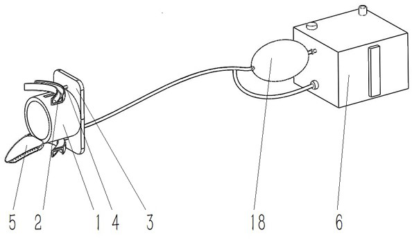
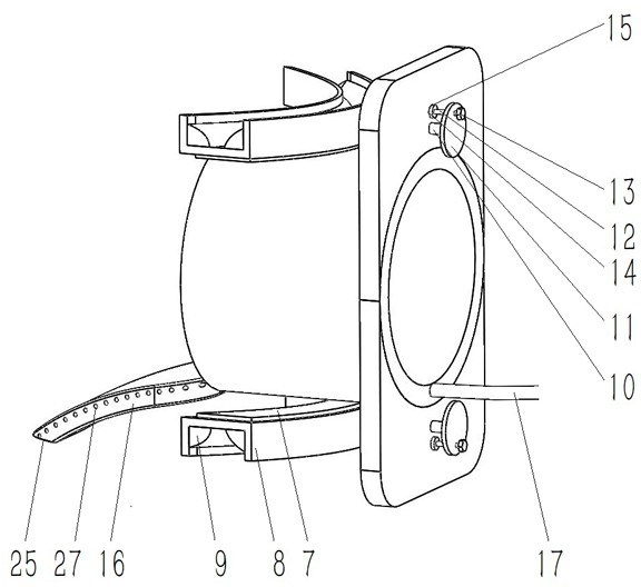
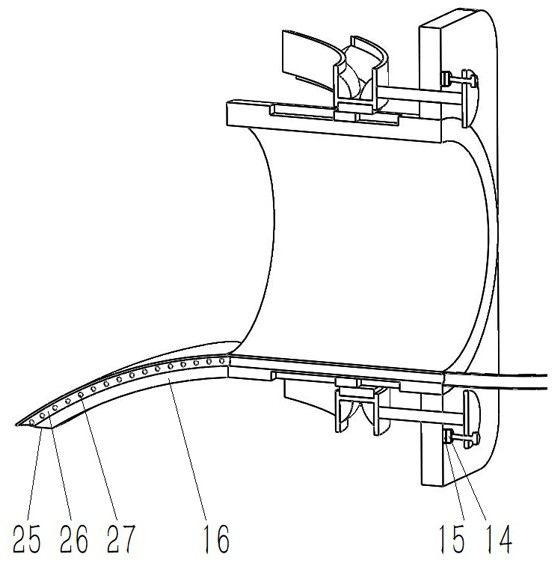
[0025] Example 1, such as Figure 1-4 shown.

[0026] An oral support device for gastroscopy. It includes a lower mirror tube 1, the lower mirror tube 1 is a hollow tubular structure, and the pipeline of the gastroscope is inserted into the patient's stomach through the lower mirror tube 1. Both upper and lower sides of the lower mirror tube 1 are provided with occlusal devices 2 for the patient to occlude. The front end of the lower mirror tube 1 is provided with a fixed plate 3, and an occlusal moving device 4 is arranged between the fixed plate 3 and the occlusal device 2 to move the front and rear positions of the occlusal device 2 to suit different patients. Teeth. The lower side of the rear end of the lower mirror tube 1 is provided with a tongue depressor 5, which can limit the tongue in the patient's mouth by the tongue depressor 5. esophagus. Reduce the discomfort when the tube passes through the throat, thereby avoiding the resistance of the patient. The tongue...

Embodiment 2

[0037] Example 2, such as Figure 4 shown.

[0038] The rest are the same, but the structure of the negative pressure assembly 18 is different.

[0039] The negative pressure assembly 18 includes a negative pressure pump 30, and the negative pressure pump 30 is powered by an external power supply. The negative pressure pump 30 is preferably a micro-gear pump of Yuanxun Intelligent Technology Co., Ltd., the model is FG300. The negative pressure pump 30 is connected with the connecting pipe 17, the negative pressure pump 30 and the inner cavity of the recovery box 19 through pipelines; the top surface of the recovery box 19 is equipped with a suction button 31, the The suction button 31 is connected with the negative pressure pump 30 through a circuit. Press the suction button 31 to control the negative pressure pump 30 to start, and suck the saliva into the recovery box 19 .

[0040] When the present invention is in use:

[0041] Unscrew the limiting handwheel, and adjust ...


PUM


Abstract

Oral cavity supporting equipment for gastroscopy comprises a lower endoscope tube, occlusion devices are arranged on the upper side and the lower side of the lower endoscope tube, a fixing plate is arranged at the front end of the lower endoscope tube, and occlusion moving devices are arranged between the fixing plate and the occlusion devices and used for moving the front-back positions of the occlusion devices. A saliva resorption device is arranged in the tongue pressing device; the device has the advantages that the occlusion device is matched with the occlusion moving device to adapt to oral cavities of different patients, on one hand, the occlusion comfort of the patients can be improved, and tooth pain caused during occlusion is avoided; on the other hand, the lower mirror tube is prevented from being bitten to damage inspection appliances; the tongue in the oral cavity of the patient can be limited through the tongue pressing device; discomfort caused when a pipeline passes through the throat is reduced, and then reaction of a patient is avoided; by means of the saliva resorption device, saliva swallowed by the patient in the examination process can be resorbed, and the saliva is prevented from flowing into the stomach to affect examination.

Description

technical field [0001] The invention relates to an oral cavity support device, in particular to an oral cavity support device for gastroscopy. Background technique [0002] Gastroscopy is a method of medical examination and also refers to the equipment used in this examination. It is inserted into the stomach through a thin, flexible tube, allowing doctors to directly observe lesions in the esophagus, stomach, and duodenum, especially for minor lesions. Gastroscopy can directly observe the real situation of the inspected part, and can further confirm the diagnosis through pathological biopsy and cytological examination of suspicious lesions. It is the preferred inspection method for upper gastrointestinal lesions. [0003] During gastroscopy, the patient's mouth needs to be stretched, so that the tube can be better inserted into the stomach, and at the same time, it is also necessary to prevent the patient from resisting the bite and damaging the examination equipment. How...

Claims


Application Information

Patent Timeline
IPC IPC(8): A61B1/273A61B1/24A61B1/00
CPCA61B1/2736A61B1/24A61B1/00131A61B1/00094A61B1/0008
Inventor 梅璐王昊阳杨明华
Owner 郑州大学第五附属医院
Learn More