Visual Colles fracture and reduction model device

A model device and model technology, which is applied in the field of visualized Colles fracture and reduction model device, can solve the problems of model fixation and inability to display the manual reduction process, etc., to achieve the effects of deepening understanding, improving the quality and efficiency of clinical work, and reducing fear and pain

Pending Publication Date: 2022-07-29
宁德市闽东医院
View PDF0 Cites 1 Cited by
  • Summary
  • Abstract
  • Description
  • Claims
  • Application Information

AI Technical Summary

Problems solved by technology

[0003] In view of the above-mentioned technical deficiencies, the purpose of the present invention is to provide a visual Colles fracture and reduction model device, which effectively s

Method used


Image

  • Visual Colles fracture and reduction model device

Examples


Embodiment 1

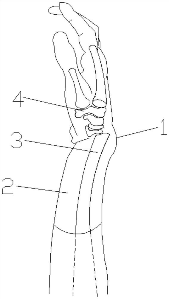
[0017] Example 1, as figure 1 As shown, a visual Colles fracture and reduction model device includes a medical upper limb forearm model 1, a visual structure 2, a distal radius internal structure model 3, and a wrist joint structure model 4. The medical upper limb forearm model is set at the distal radius of the radius. It is a visible structure, and the surface is set as an openable and closable yellow invisible soft imitation leather material; the internal structure model 3 of the distal radius and the wrist joint structure model 4 are movably connected by clues to simulate ligament wrist joint and distal radius fractures Structural model; the simulated ligament wrist joint and distal radius fracture structural model are set inside the medical upper limb forearm model 1 . The visual Colles fracture model device has soft material with good elasticity and resistance to stretching, which can be rotated and pulled, which is convenient for the demonstration of the manual reductio...


PUM


Abstract

The invention relates to the technical field of medical teaching fracture model devices, and discloses a visual Colles fracture and reduction model device which comprises a medical upper limb forearm model, a visual structure, a simulated ligament wrist joint and a distal radius fracture structure. According to the appearance, the distal radius of the medical upper limb forearm model with the silver fork-like deformity is of a visual structure made of a transparent soft skin-imitating material, and the surface of the model is made of an openable yellow invisible soft skin-imitating material; the model is internally provided with a simulated ligament wrist joint and distal radius fracture structure, and is made of a white hard plastic material; after operation reduction, fracture is reproduced according to an injury mechanism, and reutilization is realized. The visual Colles fracture model device disclosed by the invention has high simulation appearance and clinical touch hand feeling, and completely meets clinical training requirements; through the application of the model device in teaching, the problems that a model is fixed and cannot display a technique resetting process and a resetting effect are effectively solved; and the clinical work quality and efficiency are improved.

Description

technical field [0001] The invention relates to the technical field of a medical teaching fracture model device, in particular to a visual Colles fracture and reduction model device. Background technique [0002] The existing medical models are fixed and immovable. Although they can simply show the appearance of Colles fractures, they cannot show the process and effect of manual reduction, nor can they be used for teaching and training in clinical plaster casting. SUMMARY OF THE INVENTION [0003] In view of the above-mentioned technical deficiencies, the purpose of the present invention is to provide a visual Colles fracture and reduction model device, which effectively solves the problem that the model is fixed and cannot display the manual reduction process and reduction effect; at the same time, it can meet the teaching and training needs of clinical plaster casting. . [0004] In order to solve the above-mentioned technical problems, the present invention adopts the ...

Claims


Application Information

Patent Timeline
IPC IPC(8): G09B23/30
CPCG09B23/30
Inventor 邹斌缪陈芳袁翠华周东刘成招
Owner 宁德市闽东医院
Why Eureka
Try Eureka