Computer aided characteristic registration identifying method for medical homolateral fundus image

A computer-aided medical image technology, applied in the field of biomedical image processing and recognition, can solve the problems of inconvenient unification, easy fatigue, high labor intensity, etc., and achieve the effect of reducing intensity, relieving trouble and fatigue

Inactive Publication Date: 2002-11-06
SOUTH CHINA UNIV OF TECH
View PDF0 Cites 7 Cited by
  • Summary
  • Abstract
  • Description
  • Claims
  • Application Information

AI Technical Summary

Problems solved by technology

[0003] The purpose of the present invention is to overcome and solve the problem of manual fitting of fundus images in the existing fundus clinical diagnosis of ophthalmology, and the need to operate and observe under the working environment of transmitted light, which is often complicated and laborious and labor intensive. , easy fatigue, and inconvenient unification and other problems and problems such as troubles and burdens, research and invention can overcome and solve the problems and burdens of existing fundus image registration rec

Method used


Image

  • Computer aided characteristic registration identifying method for medical homolateral fundus image
  • Computer aided characteristic registration identifying method for medical homolateral fundus image
  • Computer aided characteristic registration identifying method for medical homolateral fundus image

Examples


Embodiment Construction

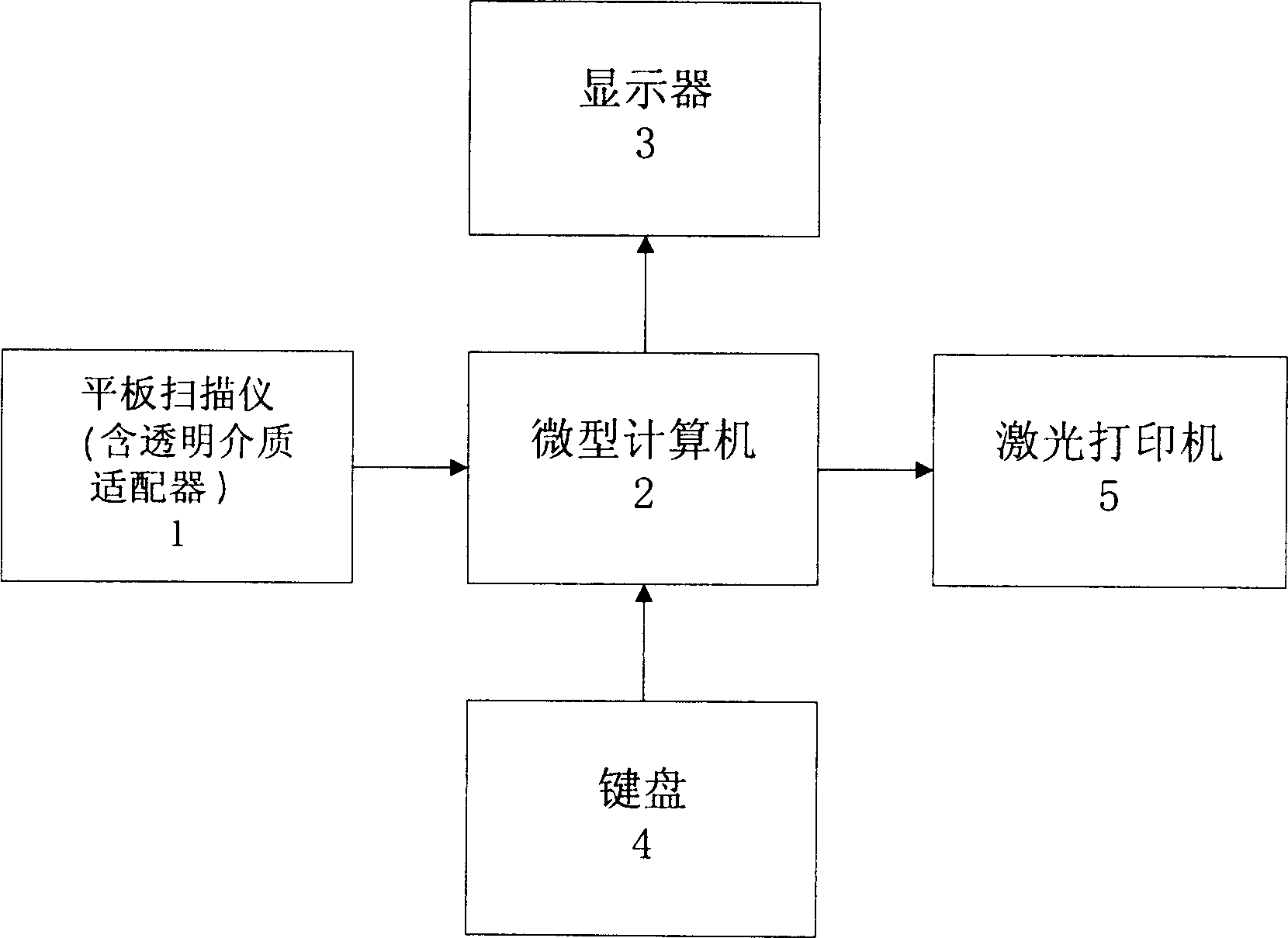
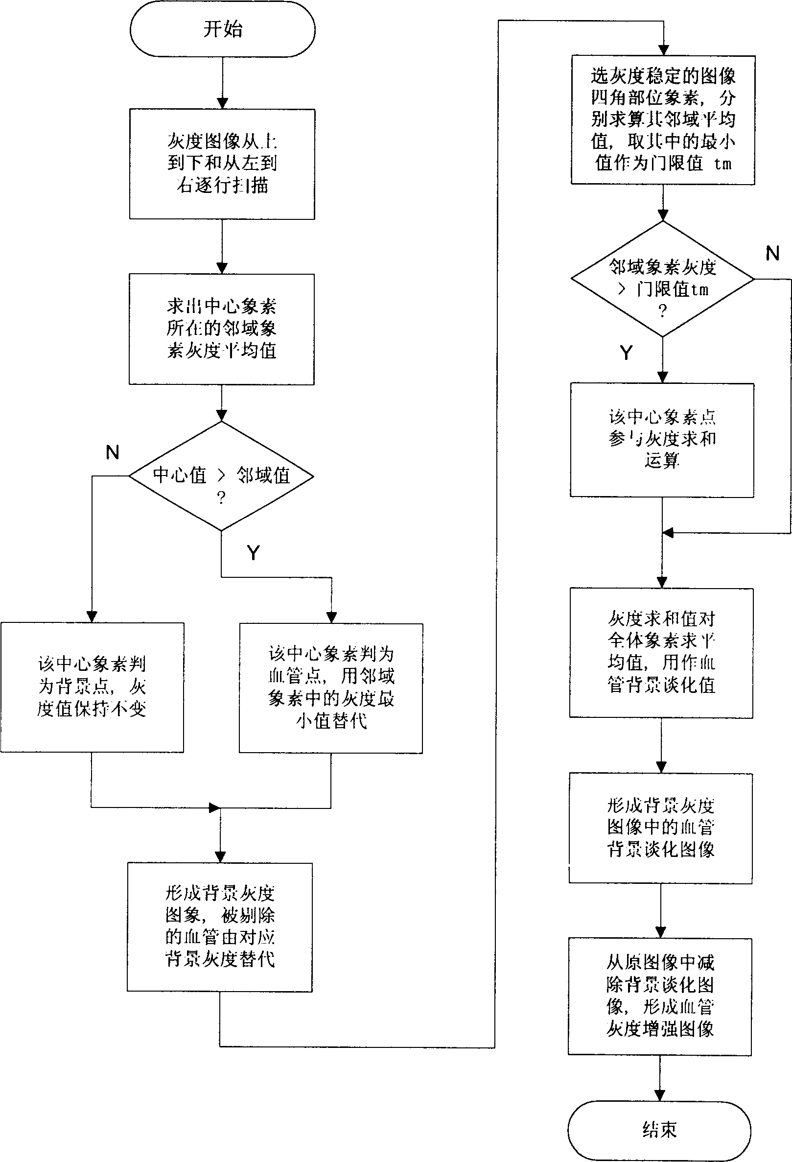
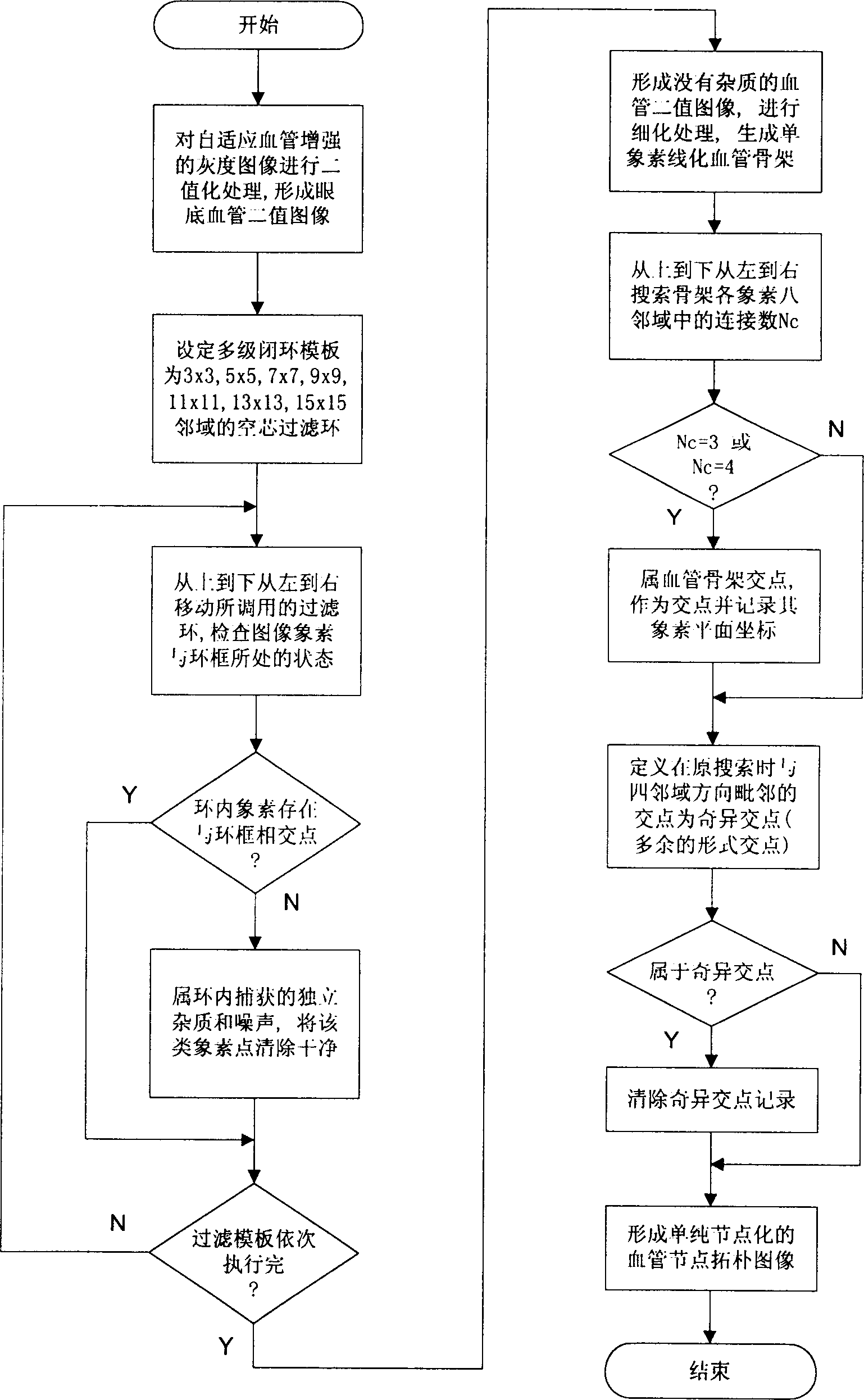
[0009] After research and testing, the inventor thinks that the optimal way to realize the present invention can be as follows: (1) the identification equipment system used in the present invention can be according to figure 1 The electrical connections shown and described in the above instructions are connected through corresponding output cables. Wherein, the flatbed scanner 1 can adopt the HP3C type, the microcomputer 2 can adopt the PIII type, the monitor 3 can adopt the flat type, and the laser printer 5 can adopt the HP series; (2) press Figure 2 to Figure 5 Shown program flowchart, can adopt C language to compile various processing procedures, and carry out step by step by each processing procedure step of the inventive method described in above description, just can implement the present invention preferably. The example diagram of the processed ipsilateral fundus medical image is as follows: the example of the original fundus blood vessel grayscale image Figure 6 A...


PUM


Abstract

The present invention relates to computer applying technology. The registration identifying system of the present invention consists of plate scanner with transparent medium adaptor, microcomputer with keyboard and display, laser printer electrically connected together via signal cables. The registration identifying process includes mainly adaptive enhancing of homolateral fundus vessel image; intersection point and pure node extraction of fundus vessel characteristic; normalization of fundus vessel characteristic nodes; and neighboring region registration and judgement of fundus vessel characteristic nodes. The computer-aided judgement means has light labor strength and other advantages.

Description

(1) Technical field: [0001] The invention relates to a computer-aided feature fitting recognition method of an ipsilateral fundus medical image, which belongs to computer application technology, and particularly relates to biomedical image processing and recognition technology. (two) background technology: [0002] Ophthalmology uses fundus cameras to take fundus photo images or negative film images during fundus clinical diagnosis or medical screening, and its archived data is gradually increasing. The left and right sides of the fundus image are different. To study or compare the follow-up photos or negatives in different periods, it is usually done manually now. The negatives need to be observed in a working environment with transmitted light. It is a very strenuous and complicated work, and the operators are labor-intensive, prone to fatigue, and inconvenient to unify and other troubles and burdens. (3) Contents of the invention: [0003] The purpose of the present in...

Claims


Application Information

Patent Timeline
IPC IPC(8): G06F19/00
Inventor 林土胜徐锦堂卢斌张小玲
Owner SOUTH CHINA UNIV OF TECH
Why Eureka
Try Eureka