Method for non-invasive clinical monitoring intracranial edema

A clinical monitoring and brain technology, applied in diagnostic recording/measurement, medical science, sensors, etc., can solve the problem of blindness in treatment, lack of data on the evolution of edema and hematoma, and inability to dynamically observe the changes of patients detached from intracerebral hematoma in real time, etc. question

Inactive Publication Date: 2003-05-28
CHONG QING BORN FUKE MEDICAL EQUIP CO LTD
View PDF0 Cites 11 Cited by
  • Summary
  • Abstract
  • Description
  • Claims
  • Application Information

AI Technical Summary

Problems solved by technology

At present, in the process of treating cerebral edema and hematoma at home and abroad, there is a lack of real-time data on the evolution of edema and hematoma, and it i

Method used


Image

  • Method for non-invasive clinical monitoring intracranial edema
  • Method for non-invasive clinical monitoring intracranial edema
  • Method for non-invasive clinical monitoring intracranial edema

Examples


example 1

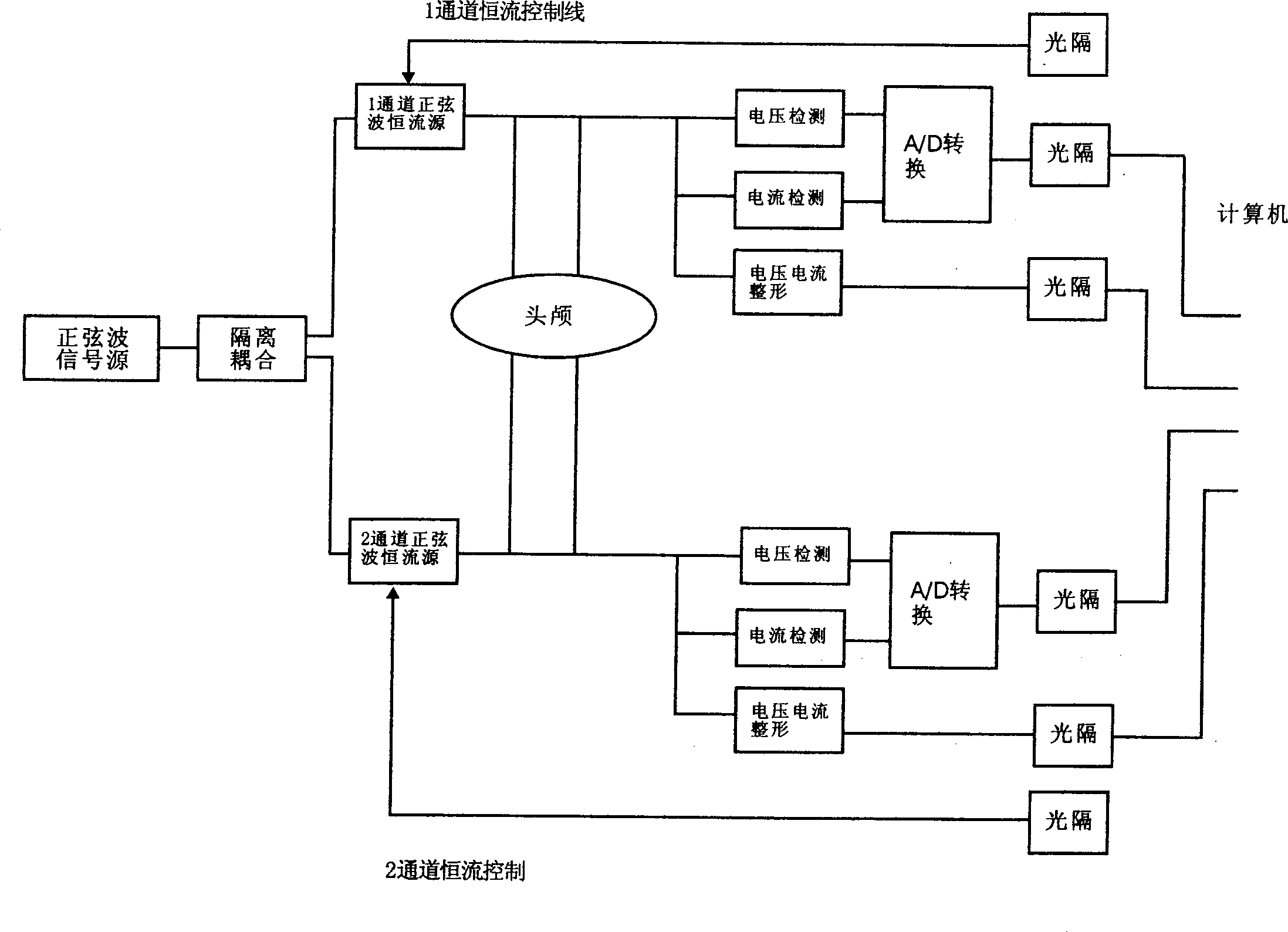
[0016] Example 1: Using the present invention, two pairs of electrodes are applied to add current from the front and back symmetrical points of the skull surface to form a current field, and the value of potential or impedance is recorded, and intracranial hematoma and edema are inferred from the value of potential or impedance.

[0017] Connection method: 16 pairs of electrodes can be arranged in an orderly manner on the surface of the skull.

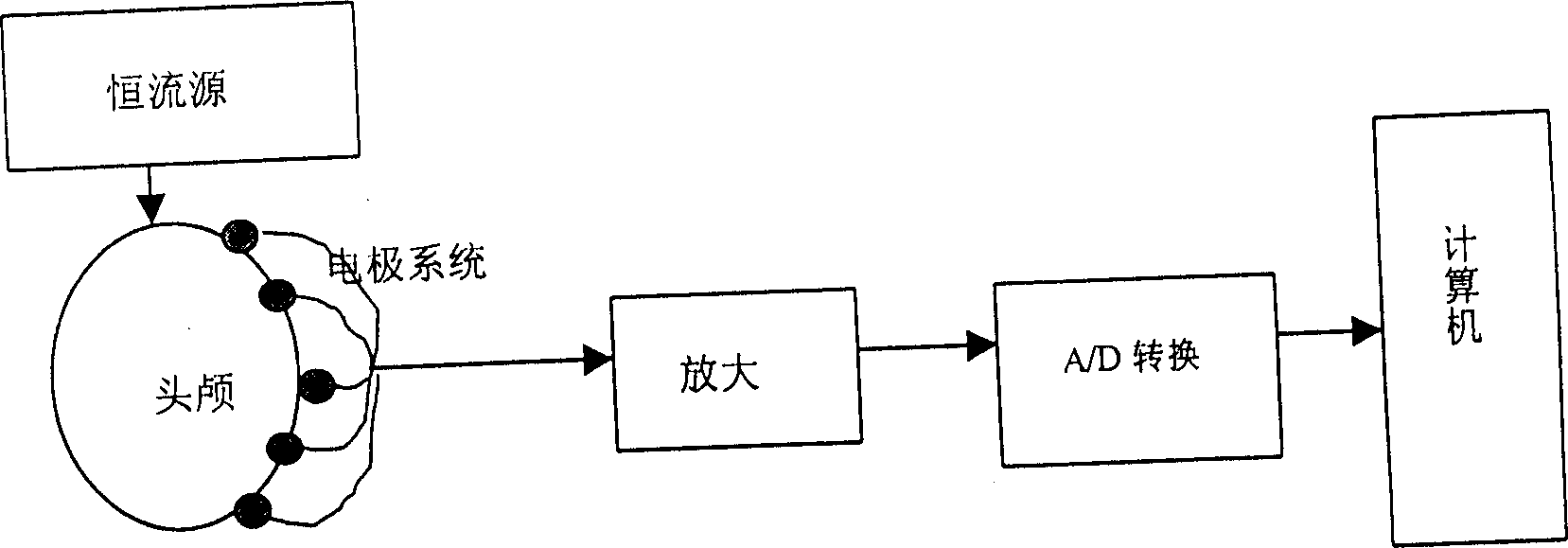
[0018] Measuring principles such as figure 2 So not.

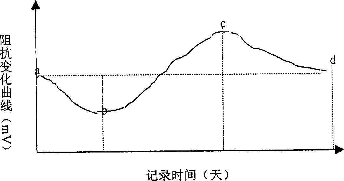
[0019] For measurement results, see image 3 , The left side first bleeds to form a hematoma, and then edema appears around the hematoma. The right side is normal. The figure shows the contrast curve of impedance change on the left and right sides.

[0020] Conclusion 1: The appearance of hematoma is manifested by the decrease of impedance on the bleeding side. Section ab in the figure.

[0021] Conclusion 2: After the bleeding stops, the impedance rises, which means edema o...


PUM


Abstract

A method for clinically monitoring the intracranial edema without wound is disclosed. A low-frequency electric current flows through the craniocerebral surface to generate a current field. When the tissue structure in the current field changes, the electric current field is redistributed to reflect the change of intracranial edema, which is detected by phase change and displayed by computer and display device.

Description

1. Technical field [0001] The invention relates to a new technology of digital medical monitoring, in particular to a method for non-invasive clinical monitoring of intracranial edema. 2. Background technology [0002] Intracranial edema is the pathological response of brain tissue to different etiologies. Correct and timely evaluation of the nature and evolution of intracranial edema is the key to the success or failure of diagnosis and treatment of many critically ill patients. Intracranial edema occurs in a completely closed special environment. The current diagnosis of deep intracranial edema is based on the accumulated clinical experience of doctors. The commonly used detection methods referred by doctors are CT, conventional magnetic resonance and diffusion-weighted nuclear magnetic resonance. CT radioactivity is strong, and patients cannot be placed under radiation for a long time, so they cannot become a routine monitoring method. Magnetic resonance imaging is clea...

Claims


Application Information

Patent Timeline
IPC IPC(8): A61B5/05
Inventor 何为熊新翔吴琪
Owner CHONG QING BORN FUKE MEDICAL EQUIP CO LTD
Why Eureka
Try Eureka