
Device and method for magnetic resonance imaging

A technology of magnetic resonance imaging and gradient magnetic field, which is applied in the direction of measurement using nuclear magnetic resonance imaging system, analysis using nuclear magnetic resonance, magnetic resonance measurement, etc.

Inactive Publication Date: 2004-05-19
TOSHIBA MEDICAL SYST CORP
View PDF1 Cites 10 Cited by
  • Summary
  • Abstract
  • Description
  • Claims
  • Application Information

AI Technical Summary

Problems solved by technology

However, since the streaming pulses are impressed on the same axis as the readout gradient field used for frequency encoding, the instantaneous space that can be left for the streaming pulses in the pulse train is limited.
Therefore in some cases flow pulses cannot be applied with optimal time integral values

Method used


Image

  • Device and method for magnetic resonance imaging
  • Device and method for magnetic resonance imaging
  • Device and method for magnetic resonance imaging

Examples


Embodiment Construction

[0030] The embodiments according to the present invention will now be described with reference to the drawings.

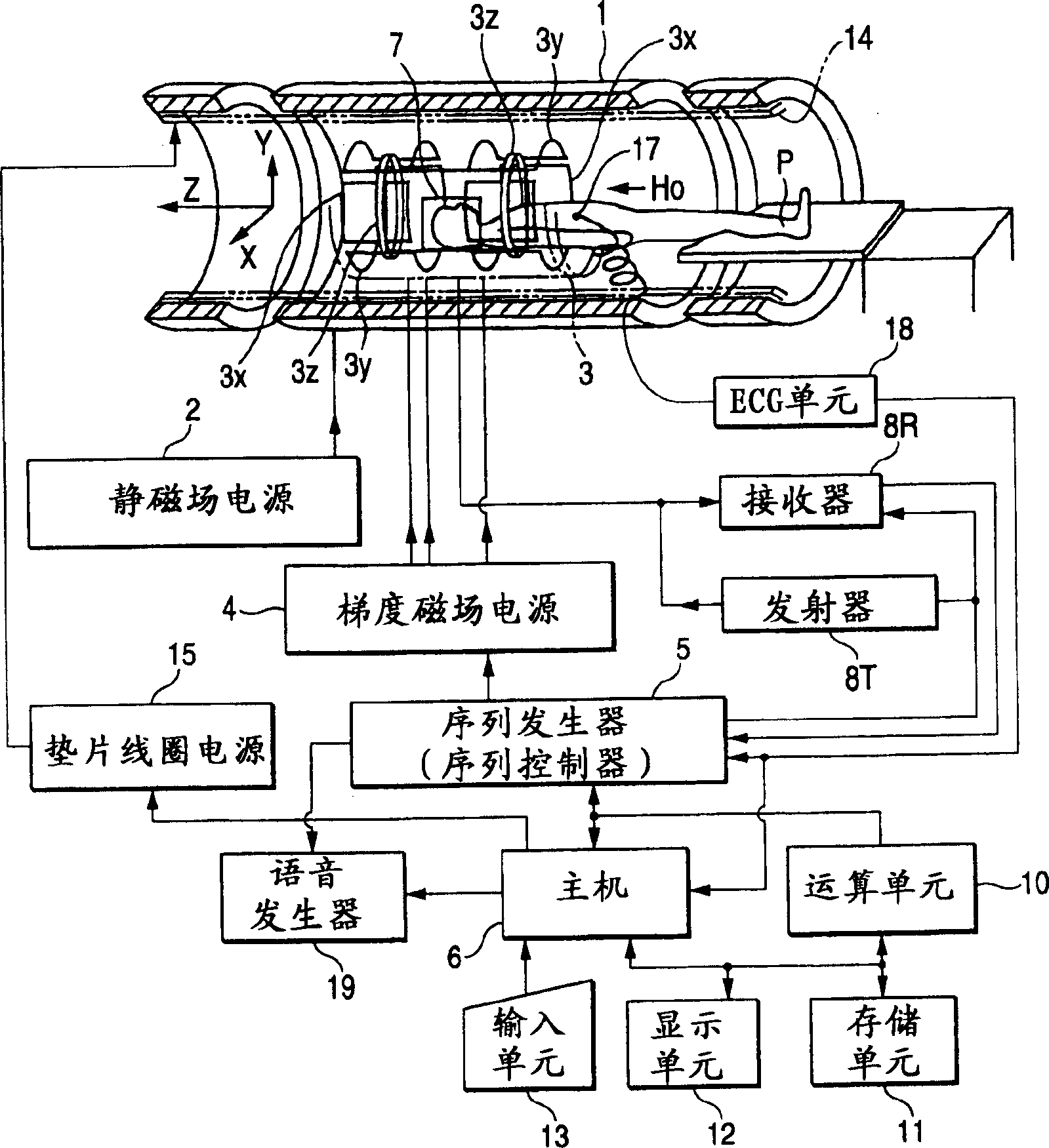
[0031] figure 1 The schematic structure of the magnetic resonance imaging apparatus according to the present embodiment is shown. The magnetic resonance imaging device includes: a part of the patient bed on which the subject patient P lies, a static magnetic field generating part that generates a static magnetic field, a gradient magnetic field generating part for adding positioning information to the static magnetic field, and transmitting / receiving RF (radio frequency) signals The transmitting / receiving part, the control / computing part that controls the entire system and image reconstruction, the electrocardiogram measurement part that measures the ECG (electrocardiogram) signal representing the heart phase signal of the patient P, and the breath-hold command part that commands the patient P to hold his breath temporarily .

[0032] The static magnetic field genera...


PUM


Abstract

A magnetic resonance imaging apparatus comprises an RF coil unit which generates RF pulses toward a subject, and which receives an MR signal from the subject, gradient magnetic field coils which generate a gradient magnetic field for slice selection, a gradient magnetic field for phase encoding and a gradient magnetic field for frequency encoding, an arithmetic unit which generates image data on the basis of the MR signal, and a sequence controller which controls the second gradient magnetic field coils in order to generate flow pulses for dephasing or rephasing a spin of a blood flow within the subject, in the same direction as that of the phase encoding gradient magnetic field.

Description

[0001] Cross-reference of related applications [0002] This application is based on and claims the priority benefit of the previous Japanese Patent Application No. 2002-295190, which was filed on October 8, 2002 and the entire content of which is incorporated by reference. Technical field [0003] The present invention relates to a magnetic resonance imaging apparatus and method, which is equipped with MR angiography capable of distinguishing arteries and veins without using any contrast medium. Background technique [0004] Magnetic resonance imaging is an imaging method in which a subject's nuclear spin in a static magnetic field is magnetically excited by its Larmor frequency radio frequency signal, and the image is reconstructed by the magnetic resonance signal generated by the excitation. In the field of magnetic resonance imaging, an imaging method that has recently begun to attract attention is the type of MR angiography that does not use any contrast medium. [0005] In M...

Claims


Application Information

Patent Timeline
Patent Type & Authority Applications(China)
IPC IPC(8): G01R33/48A61B5/055G01N24/08G01R33/563
CPCG01R33/56316G01R33/56308G01N24/08
Inventor 宫崎美津惠
Owner TOSHIBA MEDICAL SYST CORP
Learn More