Electric impedance tomographic imaging method and apparatus for imaging monitoring beside sickbed

A technology of electrical impedance tomography and imaging method, applied in the field of medical devices or instruments, can solve the problems of insufficient resolution of impedance images, no breakthroughs, and inability to accurately detect clinical lesions.

Inactive Publication Date: 2004-09-08
FOURTH MILITARY MEDICAL UNIVERSITY
View PDF0 Cites 38 Cited by
  • Summary
  • Abstract
  • Description
  • Claims
  • Application Information

AI Technical Summary

Problems solved by technology

They have explored many aspects of the clinical application of impedance imaging, and have done a lot of work on the functional imaging of the digestive system, respiratory system, brain, heart, breast, etc., but the resolution of the impedance image is not high enough. , cannot reach the level of resolution of current CT and MRI, and cannot be used for accurate detection of clinical diseases like CT and MRI, and no breakthrough has been made so far

Method used


Image

  • Electric impedance tomographic imaging method and apparatus for imaging monitoring beside sickbed
  • Electric impedance tomographic imaging method and apparatus for imaging monitoring beside sickbed
  • Electric impedance tomographic imaging method and apparatus for imaging monitoring beside sickbed

Examples


Embodiment Construction

[0059] The present invention will be further described in detail below in conjunction with the accompanying drawings and the embodiments completed by the inventor according to the technical solution.

[0060] 1. Overall structure

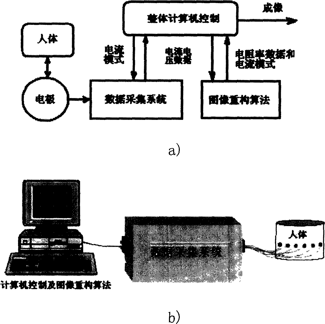
[0061] The overall structure of the present invention is as figure 1 As shown, a) in the figure is a block diagram, and b) is a schematic diagram of the device.

[0062] Overall computer control: control the data acquisition system and the algorithm for reconstructing the image, provide an interface for the user, and enable the staff to control the process of image monitoring.

[0063] Electrodes: Using ECG electrodes or EEG electrodes, the excitation current is applied to the human body, and the signals of the human body are transmitted to the data acquisition system.

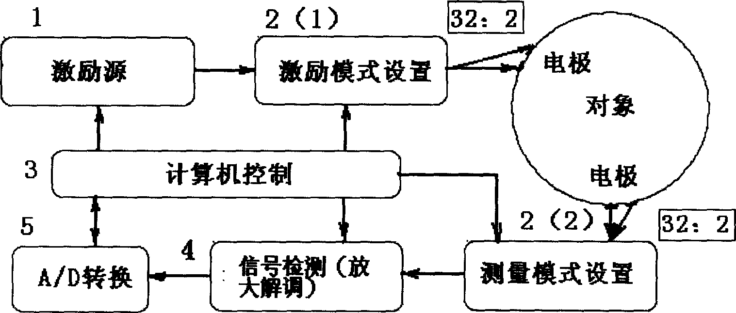
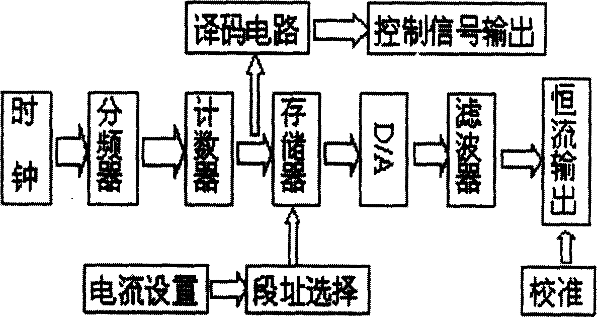
[0064] Data acquisition system: Provide AC constant current excitation, high-precision signal detection, program-controlled setting of drive measurement mode, etc.

[0065] Image r...


PUM


Abstract

The present invention discloses electric impedance tomographic imaging method and apparatus for imaging nursing beside sickbed. The integral computerized data collecting system and the re-construction algorithm provides the interface and controls the imaging nursing process. The apparatus includes one computer and one connected data collecting system, the computer is equipped with program for controlling the data collecting system and algorithm for re-constructing image, and the data collecting system has also control module to apply excitation current to body signal collecting electrodes. The present invention features the several pairs of electrodes for applying excitation current and collecting body signals, the data collecting system providing constant AC excitation, high precision signal detection and programmed driving and measuring mode, and the calculation of body resistivity distribution and image display.

Description

technical field [0001] The invention belongs to medical equipment or instruments, and relates to an electrical impedance tomography method and a device thereof which can be used for bedside image monitoring. Background technique [0002] Some diseases with high morbidity in clinical diagnosis and treatment now require imaging technology that can be used for bedside monitoring: stroke, subacute traumatic brain injury, chronic intracranial hemorrhage, postoperative brain complications, neonatal intracranial hemorrhage, Delayed internal hemorrhage or local ischemia of abdominal organs, organ transplantation, etc. all require bedside image monitoring technology. [0003] However, none of the existing imaging technologies can be used for bedside image monitoring: such as the existing CT, MRI, PET, ultrasound, etc., which not only require huge equipment and high costs, are complicated to operate, have certain damage to the human body, and are impossible Do bedside dynamic, real-t...

Claims


Application Information

Patent Timeline
Patent Type & Authority Applications(China)
IPC IPC(8): A61B5/053
Inventor 董秀珍付峰尤富生刘锐岗史学涛
Owner FOURTH MILITARY MEDICAL UNIVERSITY
Why Eureka
Try Eureka